Review Article - Imaging in Medicine (2020) Volume 12, Issue 3
Nuclear Medicine in Sports Medicine
Margarita Pagou* & Vasileios PoulantzasAsklipios Medical Diagnostic Centre, Athens, Greece
- Corresponding Author:
- Margarita Pagou
Asklipios Medical Diagnostic Centre
Athens, Greece
E-mail: marpagou@otenet.gr
Abstract
Nuclear medicine imaging has applications in the diagnosis and monitoring of bony and soft tissue sports related injuries. Skeletal scintigraphy may localize traumatic disorders. Dynamic studies show changes in many conditions such as bone stress, fracture, periosteal reaction, osteochondritis dissecans, enthesopathy, osteonecrosis, complex regional pain syndrome, degenerative change. Special views can be used in fracture, periosteal reaction, osteochondritis dissecans, enthesopathy and abnormalities in the spine. Magnification techniques have also been described. Pinhole imaging can detect fracture, osteonecrosis, impingement syndromes. SPECT imaging may give valuable information in the detection of fracture, avulsion injury, joint injuries, osteonecrosis, back pain and rhabdomyolysis. SPECT/CT allows detection of osteochondritis dissecans, enthesopathy, joint injuries, impingement syndromes and abnormalities in the spine. Lymphoscintigraphy is a diagnostic tool. There is interest in PET studies at traumatic brain injury and at the neurotransmitter level. PET/CT can be useful in selected athletic injuries.
Keywords
scintigraphy â– special views â– pinhole views â– SPECT â– SPECT/CT â– PET â– PET/CT â– sports injuries
Bone stress
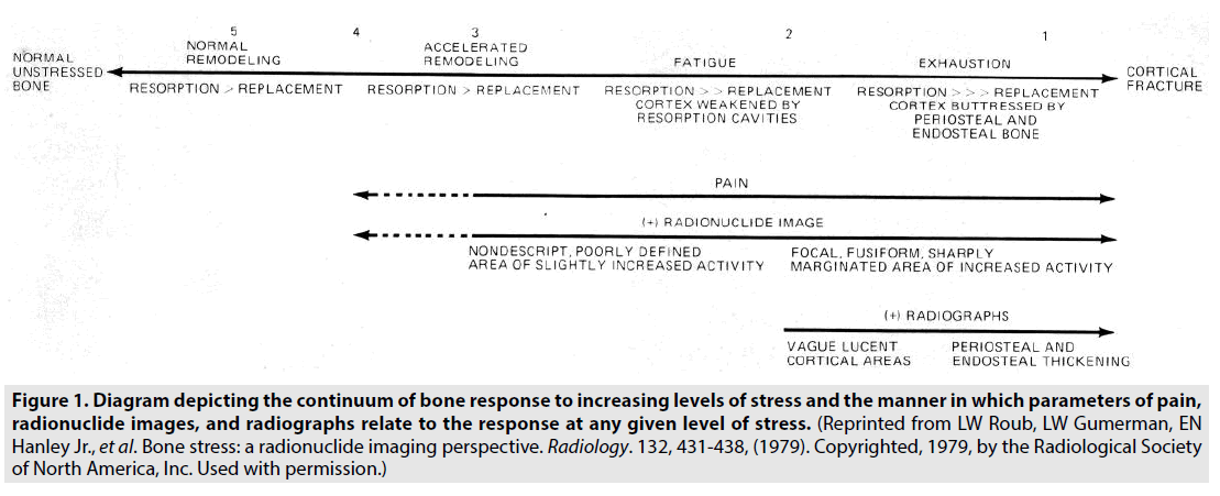
Bone stress can eventually lead to the fracturing of bone. A schema (FIGURE 1) has been proposed to explain the changes in bone in response to repetitive stress and the occurrence of positive radionuclide images and negative radiographs in the same patient with bone stress [1].
Figure 1: Diagram depicting the continuum of bone response to increasing levels of stress and the manner in which parameters of pain, radionuclide images, and radiographs relate to the response at any given level of stress. (Reprinted from LW Roub, LW Gumerman, EN Hanley Jr., et al. Bone stress: a radionuclide imaging perspective. Radiology. 132, 431-438, (1979). Copyrighted, 1979, by the Radiological Society of North America, Inc. Used with permission.)
Most femoral stress injuries are associated with running [2].
Bone stress reactions in ballet dancers, most prevalent in the tibiae, have been demonstrated by a diffusely increased uptake of a radiotracer [3].
In patients with stress changes or fractures of the sesamoid bones at the head of the first metatarsal, three-phase skeletal scintigraphy can give objective evidence of the exact site of the abnormality [4].
Fracture
â– Stress fracture
Stress fracture is an overuse injury caused by muscle forces combined with bending and impact forces acting on the bone, which has not adapted to loading [5]. Repeated application of stress lower than the stress required to fracture the bone in a single loading results in a partial or complete stress fracture [6].
The minimum time for skeletal scintigraphy to become abnormal following a fracture is agedependent; however, after injury, 80% of all fractures can be abnormal by 24 hours, and 95% by 72 hours [7].
If scintigraphy is performed in the acute phase within 4 weeks, the flow and blood pool images show increased activity [8].
Skeletal scintigraphy may be positive 10 to 14 days prior to the appearance of any findings on plain radiographs. MRI is comparable in terms of sensitivity, but within 24 to 48 hours of injury, skeletal scintigraphy may not be as sensitive as MRI [9].
The usefulness of MRI imaging of the hip and adjacent bony structures has been reported [10]. The impression is that the accuracy of skeletal scintigraphy for the diagnosis of femoral neck stress fractures approaches 100% if oblique views of the pelvis are taken, and then the uptake anterior and posterior to the hip is projected away from the femoral neck and false positives are avoided [11]. Pinhole imaging in the hips has also helped to identify the pattern of stress fractures in the inferior aspect of the femoral neck, which is the typical site of occurrence in athletes [12]. Also, to increase the sensitivity of skeletal scintigraphy for the detection of upper femoral lesions, frog-leg views should be obtained in addition to the standard anterior view (FIGURE 2) [13].
Figure 2: Position for frog-leg view of right hip. (Reprinted from W Ammann, J Matzinger, DR Lloyd-Smith, et al. Femoral stress abnormalities: improved scintigraphic detection with frog-leg view. Radiology. 169, 844-845, (1988). Copyright © RSNA, 1988. Used with permission.)
Lateral blood pool imaging in patients with suspected stress fractures of the tibia could potentially have clinical relevance depending on severity and prognosis [14].
The plantar projection of the feet, which clearly shows the individual structures of the metatarsus and phalanges with minimal overlap, allows stress fractures and degenerative or posttraumatic joint disease, sesamoiditis, ligament avulsion injuries or tendinitis to be detected with ease. Medial and lateral views of the feet also allow the posterior half of the tarsus to be clearly visualized for most conditions of the calcaneum, such as fractures, bruising, enthesopathy, and bursitis. Clear identification of fractures through the lateral and intermediate cuneiform bones, as well as the post-traumatic change in the third tarsometatarsal articulation, may be seen in the magnified anterior oblique view [12].
Skeletal scintigraphy in pediatric athletes can be helpful for the identification of stress fractures in long bones, complex regional pain syndrome, back pain and other causes of pain [15].
Persistent and increasing bone pain during demanding physical activity, even in the presence of a prior normal skeletal scintigraphy, may represent stress fracture and a repeat skeletal scintigraphy may be needed [16].
Stress fractures are associated with a plethora of sports. For example, fractures of the lumbar spine are associated with ballet, running, and gymnastics, fractures of the humerus and ulnar shafts with baseball, and fractures of the posteromedial olecranon with throwing sports [17]. The most common stress fractures in athletes, most frequent from running, were found in the tibia, followed by fractures in the tarsals, metatarsals, femur, fibula, pelvis, sesamoids and spine [18].
Although sensitive for acute fractures, skeletal scintigraphy is non-specific and may be positive in other scenarios including tumor, infection, and arthritis. The correlation of the patient’s history, physical exam, plain radiographs, and three-phase skeletal scintigraphy will assist in the correct diagnosis [19].
â– Occult fracture
An occult fracture is a fracture that is not diagnosed on plain radiography for a variety of reasons, e.g., complex regional anatomy and subtle fatigue fracture [20,21].
Of particular concern is the identification of scaphoid fracture due to the high incidence of osteonecrosis [19]. It usually results from a fall on the outstretched hand, which is often not revealed by radiographs after a delay of 7–10 days if it is a non-displaced fracture [22]. Highresolution scintigraphy and prone views of the wrists with ulnar deviation may precisely localize the scaphoid fracture [23]. For the diagnostic management of suspected scaphoid fracture, an analysis of the incremental costs incurred to save one non-union showed that the use of skeletal scintigraphy in patients with negative initial radiographs costs approximately onethird of the price of using repeated radiography up to 6 weeks after injury and is, therefore, more cost-effective [24]. Another frequent occult fracture is the hamate fracture, which is commonly associated with an injury produced while swinging a tennis racquet, baseball bat or golf club where the butt of the handle strikes the hook of the hamate and fractures it [22].
Stress fractures of the ribs in amateur golfers have been diagnosed by skeletal scintigraphy. The initial chest radiographs, including special views focusing on the ribs, were all negative [25].
In a multifactorial analysis for the early detection of fractures of the proximal femur (hip) with skeletal scintigraphy and normal or equivocal radiographs, the sensitivity was 0.978 [26].
Three-phase skeletal scintigraphy with SPECT can demonstrate occult knee fractures with normal radiographs and normal overlying articular cartilage at arthroscopy [27].
For ankle and hind-foot pain undiagnosed on routine plain radiography, skeletal scintigraphy of the talus can demonstrate the specific talar joint that is injured, and tomography can confirm the presence of an osteochondral lesion [28]. A magnified posterior view of the ankle can also increase the accuracy of osteochondral injury detection of the talar dome and tibial plafond by 25% [12]. For the detection of osteochondral talar dome fractures and no abnormality on plain radiographs, CT cannot be used routinely due to its high cost. Skeletal scintigraphy appears to be a good screening procedure that will identify patients who are likely to benefit from CT [29]. Pinhole SPECT and pinhole planar imaging have been used to delineate fracture in the central talar body [30].
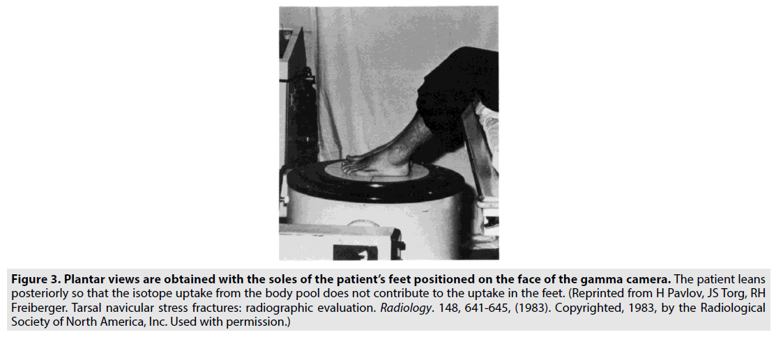
The diagnosis of a tarsal navicular stress fracture, which is only rarely evident on routine radiographs or standard tomograms, may require skeletal scintigraphy with plantar views (FIGURE 3) or anatomic anteroposterior tomograms [31].
Figure 3: Plantar views are obtained with the soles of the patient’s feet positioned on the face of the gamma camera. The patient leans posteriorly so that the isotope uptake from the body pool does not contribute to the uptake in the feet. (Reprinted from H Pavlov, JS Torg, RH Freiberger. Tarsal navicular stress fractures: radiographic evaluation. Radiology. 148, 641-645, (1983). Copyrighted, 1983, by the Radiological Society of North America, Inc. Used with permission.)
Lisfranc injury of the mid-foot includes any combination of fracture, ligament disruption, and joint dislocation [22,32,33]. Skeletal scintigraphy is a sensitive method showing minor metabolic and blood flow changes when other imaging modalities demonstrate normal results. Additionally, it may show increased uptake in the region of injury for up to 1 year after the injury [32,34]. Weight-bearing radiographs are sensitive [35,36]. A CT or MRI may be of benefit [36].
A bone bruise or bone contusion is another kind of occult fracture, in which skeletal scintigraphy shows an abnormal tracer uptake that reflects the underlying trabecular damage or microfractures with very minor cortical disruption and a reparative bone reaction, which is the speculated basis of this lesion.
Of additional interest is the ability of a three or two-phase skeletal scintigraphy to evaluate possible complications in the fracture union, as well as other complications such as avascular necrosis, complex regional pain syndrome and post-traumatic arthritis.
Moreover, skeletal scintigraphy, because of its ability to image the whole body, enables the possible cause of a referred pain to be determined, demonstrating more distant abnormal tracer uptake foci, which may represent the origin of this pain. Likewise, it permits the detection of multiple lesions when multiple skeletal injuries are suspected as well as unsuspected traumatic pathology [22].
â– Periosteal reaction (shin splints, thigh splints, ulnar splints)
This condition is thought to result from increased stress from the muscles or connective tissues on the superficial portion of the bone cortex, leading to increased bone metabolism [37].ËShin splints Ì has been defined as Ëpain and discomfort in the leg from repetitive running on hard surfaces, a forcible use of the foot flexors; diagnosis should be limited to musculotendinous inflammation excluding fracture and ischemic disorders Ì [38].
For an acute periosteal reaction in the tibia, similar sensitivity and specificity may be expected from three-phase skeletal scintigraphy and MRI. No abnormal findings may be seen on plain radiography [39]. Scintigraphic patterns can be used to differentiate periosteal reaction from stress fractures or other conditions causing pain in the lower leg in athletes. Obtaining both lateral and medial views is crucial [40].
The process is most commonly found in the tibia. The clinical equivalent in the thigh and ulna has also been described [37,41].
â– Osteochondritis dissecans (transchondral fracture)
This entity is characterized by a segment of cartilage that, together with subchondral bone, separates from an articular surface. The separation of the bony fragment may be partial or complete [42].
Repetitive throwing is considered to be one of the main etiological factors of osteochondritis dissecans of the capitellum. During the acceleration phase of throwing, the elbow joint may be stressed into a valgus position, and the capitellum may be subjected to compression and shear forces [43]. The acute flexion view of the elbow is the optimal imaging position to visualize an injury of the capitellum with skeletal scintigraphy [44].
Joint scintigraphy is valuable in the management of osteochondritis dissecans of the femoral condyles because of its superior sensitivity to changes in the lesion activity [45]. With planar views, focal hyperemia and intense radionuclide accumulation are seen in the site of the subchondral lesion with no uptake in avascular bone fragments [37]. The special intercondylar view, performed with the knee partially flexed to approximately 25 degrees and the camera positioned perpendicular to the tibial plateau, is helpful in the evaluation. Using this view, the area of involvement is seen without overlap, confirming the characteristic site and uptake of this process [46].
A potential clinical application of SPECT/CT in the knee in cases of delayed and suspected nonunion after surgery is the assessment of the surgeon regarding healing and integration of the osteochondral fragment and the decision whether the patient should be treated conservatively or re-operated on. In osteochondritis dissecans of the talus SPECT/CT also seems to be particularly helpful [47].
â– Avulsion injury
The ability of scintigraphy to reflect changes in bone physiology readily permits early identification of injuries such as avulsion lesions, stress fractures, and enthesopathies, the severity of which may not elicit secondary bone marrow edema, which is detectable by MRI. This is most pertinent for high-level athletes, for whom early diagnosis can prevent more serious sequelae [12].
An unusual avulsion of the L4 spinous process in a professional rugby player has been described. SPECT and CT showed the lesion. Plain radiography and MRI did not reveal the injury [48].
Skeletal scintigraphy showed increased uptake in an avulsion of the pubic ramus in a swimmer. Radiographs of the pelvis were negative [49]. Bone SPECT images also gave information in the evaluation of avulsion injury to the infrapatellar tendon insertion in the anterior tibia in active asymptomatic soccer players [50]. Damaged anterior cruciate ligaments were also identified with SPECT, occurring when there was avulsion of the tibial attachment in patients with acute knee pain [51].
â– Enthesopathy
Enthesopathy is a pathologic process at entheses, the sites of tendon, ligament or articular capsule firm attachment to bone [52].
Around the elbow, both lateral epicondylitis, which results from tennis, and medial epicondylitis, which results from golf, may be imaged using three-phase skeletal scintigraphy [53]. Epicondylitis of even a mild degree can be appreciated better in a magnified view of the elbow flexed to 1100 with the posterior humeral aspect placed on the face of the camera [54].
Osteitis pubis has resulted from a variety of sports activities. The use of skeletal scintigraphy in the appropriate clinical setting aids the diagnosis [55,56].
Bone SPECT images with a standard reference set of cross-sectional MRI/CT images make it much easier to identify enthesopathies and other soft tissue abnormalities of the knee, such as tendon injuries and bursitis, and also other internal derangements, such as meniscal injuries, ligament tears or strains, osteochondral injuries, bone bruising and fractures [12].
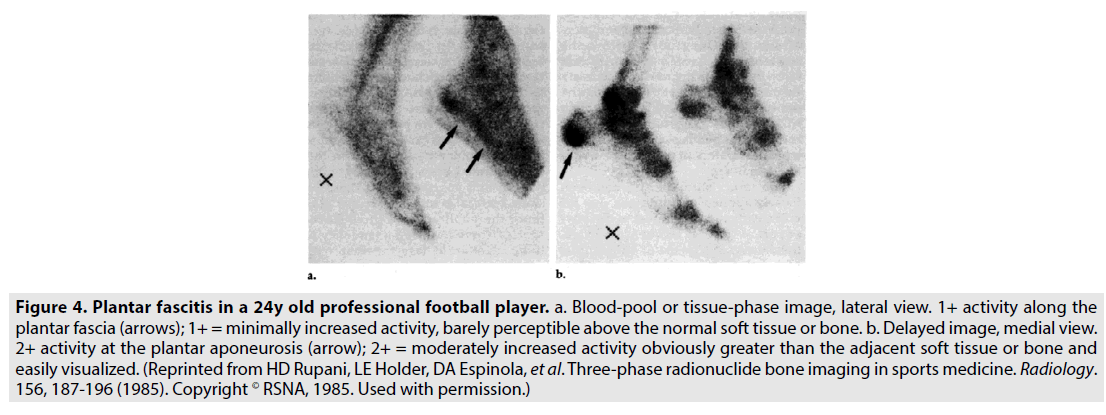
A case of injury of the insertion of the iliotibial band in an endurance athlete, where the threephase skeletal scintigraphy was positive and MRI negative, has been described [57]. Plantar fasciitis and enthesopathy at the site of insertion of the long tendinous plantar aponeurosis into the calcaneal base have also been related to sports injuries and may be elucidated by planar three-phase skeletal scintigraphy (FIGURE 4) or SPECT/CT [4,58].
Figure 4: Plantar fascitis in a 24y old professional football player. a. Blood-pool or tissue-phase image, lateral view. 1+ activity along the plantar fascia (arrows); 1+ = minimally increased activity, barely perceptible above the normal soft tissue or bone. b. Delayed image, medial view. 2+ activity at the plantar aponeurosis (arrow); 2+ = moderately increased activity obviously greater than the adjacent soft tissue or bone and easily visualized. (Reprinted from HD Rupani, LE Holder, DA Espinola, et al. Three-phase radionuclide bone imaging in sports medicine. Radiology. 156, 187-196 (1985). Copyright © RSNA, 1985. Used with permission.)
â– Joint injuries
In some sports, such as skiing, the number of joint injuries that occur is almost twice that of any other type of injury. Nuclear medicine procedures can be used to diagnose many acute injuries and most of the chronic joint abnormalities that arise in later years. The degree of metabolic activity in a joint, assuming that blood flow is maintained, is a reasonable guide to the activity of the arthritic process at any given time [59].
SPECT is very accurate, easy to perform and cost-effective. Additionally, it may give valuable information before MRI studies in the detection of meniscal tears and may be used successfully when MRI imaging is unavailable, unsuitable, inconclusive or conflictive with other clinical data. However, SPECT does not provide the anatomical detail that can be obtained with MRI [50,60]. Although the use of SPECT for the identification of meniscal damage yields a positive predictive value of 83%, the more useful clinical application in the investigation of patients presenting with acute knee pain lies in the negative predictive value of 91% [51]. Perhaps the major potential role that SPECT and planar skeletal scintigraphy offer are discriminating between potentially surgerydemanding lesions and minor injuries/healthy knees where arthroscopy could be avoided. In patients in whom SPECT is positive, recognition of the pattern of abnormality is of major assistance during arthroscopy [51,61]. SPECT/CT, PET/CT, and MRI can be used to assess patients after anterior cruciate ligament reconstruction [62,63].
Skeletal scintigraphy of the foot may be useful in sportsmen with capsular or ligamentous damage [64].
â– Osteonecrosis (avascular necrosis, bone infarction)
Osteonecrosis may be secondary to trauma [65].
By identifying a photopenic defect that is not evident on planar views, SPECT can contribute to the diagnosis of osteonecrosis of the femoral head [66].
In stage I osteonecrosis of the knee, radiographs are normal; at the same time, three-phase skeletal scintigraphy is already markedly abnormal [42].
In the fairly uncommon clinical entity of osteonecrosis of the sesamoid, three-phase skeletal scintigraphy contributes towards an early and accurate diagnosis [67,68]. Pinhole imaging is also useful in tiny bones such as tarsal navicula [69].
â– Impingement syndromes
Anterior ankle impingement can be caused by exostosis on the dorsal region of the tibiotalar joint, which may occur in soccer players (due to local and repeated microtraumas) at the cranial aspect of the talus and the navicular bone and at the anterior portion of the tibial surface [70]. Impingement exostoses, which form on strong feet, may be compatible with dancing for many years. Impingement is also found in young dancers with long, slender, flexible feet [71].
Posterior ankle impingement, caused by the same biomechanical mechanisms as the anterior one, is reported in dancers. This syndrome involves the posterior tubercle of the talus and the os trigonum, when it is present [70].
Knee impingement syndromes are very frequently reported in both professional and amateur sportsmen [72].
Radiography may be adequate for the diagnosis [71,73]. Skeletal scintigraphy and pinhole views can help [58,73,74,75]. SPECT/CT and MRI allow a proper and correct diagnosis [58,70,72]. Preoperative ankle SPECT/CT may also be helpful to clinically correlate the visual-analogscale pain score in the pre and postsurgical periods [76].
â– Complex regional pain syndrome I (Reflex Sympathetic Dystrophy)
This condition most commonly involves an extremity following even minor trauma. Three-phase skeletal scintigraphy remains a valuable imaging adjunct to clinical diagnosis. It can also be used to follow disease courses and potential treatment responses. It has significantly better sensitivity and negative predictive value than MRI and radiography in ruling out this disorder [77,78,79].
â– Back pain
Spondylolysis, which is higher in athletes who repeatedly have to hyperextend and rotate their lumbar spine, may be a cause of low back pain [80]. This injury may arise within gymnasts, javelin throwers, weightlifters, wrestlers, divers, ballet dancers, swimmers, and tennis players. Cricket and soccer are also associated with this bone stress response [80-83].
In the lumbar spine, skeletal scintigraphy with SPECT is ideal to diagnose the presence and activity of a spondylolysis lesion followed by limited computed tomography if scintigraphy is positive [84,85]. SPECT 1800 rotation posteriorly around the patient provides little to no reduction in resolution while reducing examination time by half. This 1800 acquisition can also be useful in studying obese individuals in whom the anterior data of a 3600 acquisition may be poorly attenuated [86].
When spondylolysis or spondylolisthesis is the cause of the low back pain, defects in the pars interarticularis are frequently associated with increased scintigraphic activity. SPECT imaging helps the clinician decide on whether surgery should be performed and when an athlete can return to competition. Normal skeletal scintigraphy in the presence of a radiographical pars fracture is consistent with an old, healed process, and the pars defect is unlikely to be the cause of the low back pain [87].
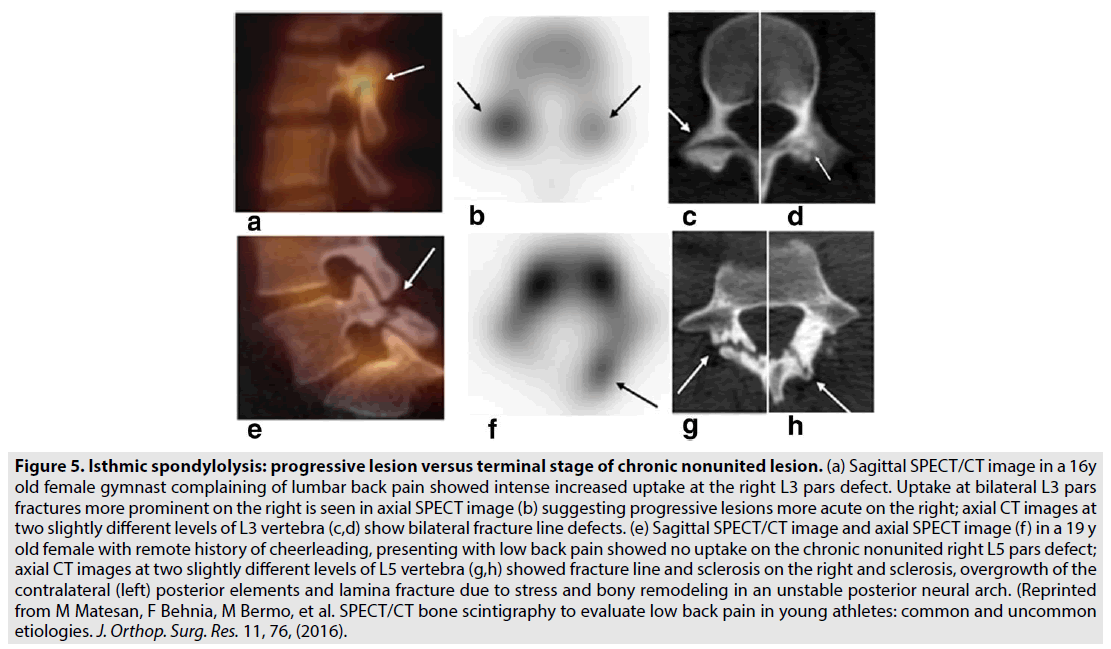
In young athletes with low back pain, SPECT/ CT skeletal scintigraphy has been shown to be useful in the detection of spondylolysis (FIGURE 5) and other common and uncommon etiologies [88]. Also, pediatric athletes with low back pain and unilateral tracer uptake on SPECT at the pars interarticularis but no defect on radiographs have been evaluated [89]. PET/CT can also be valuable in evaluating the cause of back pain in young athletes early in the disease process [90].
Figure 5: Isthmic spondylolysis: progressive lesion versus terminal stage of chronic nonunited lesion. (a) Sagittal SPECT/CT image in a 16y old female gymnast complaining of lumbar back pain showed intense increased uptake at the right L3 pars defect. Uptake at bilateral L3 pars fractures more prominent on the right is seen in axial SPECT image (b) suggesting progressive lesions more acute on the right; axial CT images at two slightly different levels of L3 vertebra (c,d) show bilateral fracture line defects. (e) Sagittal SPECT/CT image and axial SPECT image (f ) in a 19 y old female with remote history of cheerleading, presenting with low back pain showed no uptake on the chronic nonunited right L5 pars defect; axial CT images at two slightly different levels of L5 vertebra (g,h) showed fracture line and sclerosis on the right and sclerosis, overgrowth of the contralateral (left) posterior elements and lamina fracture due to stress and bony remodeling in an unstable posterior neural arch. (Reprinted from M Matesan, F Behnia, M Bermo, et al. SPECT/CT bone scintigraphy to evaluate low back pain in young athletes: common and uncommon etiologies. J. Orthop. Surg. Res. 11, 76, (2016).
Degenerative change of the spine is often exacerbated by sports activity, and the threephase skeletal scintigraphy provides significant information [4]. In facet joint arthritis the high negative predictive value of high spatial resolution SPECT images allows radionuclide skeletal imaging to be used to investigate patients with spinal pain to undergo direct facet joint injection to the correct level [91,92]. Oblique SPECT sections allow for easier differentiation between the facet and pars interarticularis abnormalities, as well as improved imaging of hips and sacrum [93].
In the thoracic spine standard planar, SPECT, pinhole and planar prone magnified images have been described.
In the cervical spine, most abnormalities of concern are in the zygapophyseal or facet joints in any sport that involves rotational movement or loading of the cervical spine, such as rugby. With SPECT and multi-head gamma cameras, high-count tomographic images are readily achievable [12].
â– Rhabdomyolysis
Overuse is the most typical cause of rhabdomyolysis associated with extreme exercise. Various types of athletes may complain of muscle damage such as long-distance runners, hurdlers, football players and weightlifters. The individual muscles which are involved can often be well-delineated by planar imaging. However, the use of SPECT imaging has helped in locating which individual muscle groups are injured [59].
â– Uncommon injuries
Accessory calcaneus described in a recreational basketball player can be well demonstrated on SPECT/CT [94].
In the assessment of a painful os intermetatarseum in a soccer player, skeletal scintigraphy was a useful diagnostic tool [95].
â– Lymphatic abnormalities
Lymphatic abnormalities related to intensive bicycling have been seen by three-phase lymphoscintigraphy of the legs [96]. A case of traumatic injury at hockey has also been described with lymphoscintigraphy [97].
â– Brain function and physical exercise
PET technology holds promise in managing sports-related concussions [98]. Nevertheless, there is no unique PET or SPECT “profile” that has been clinically validated with traumatic brain injury [99].
PET ligand activation studies at the neurotransmitter level will potentially aid clinical applications of endurance training, both as a preventative or therapeutic intervention [100].
Discussion
In the present review, a variety of sports injuries are presented. Minor, mild or significant pain, as well as other symptoms, contribute substantial information in the diagnosis (TABLE 1).
| Bone stress: Dull pain; exercise-induced pain; tenderness |
| Stress fracture: Pain at rest; exercise related pain relieved by rest; persistent and increasing pain; tenderness; swelling; cramping sensation; deformity |
| Occult fracture: Pain; tenderness; swelling |
| Periosteal reaction: Acute and chronic pain; intermittent pain |
| Osteochondritis dissecans: Slight pain; pain usually aggravated by activity and relieved by rest |
| Avulsion injury: Intense and sudden pain; swelling; bruising |
| Enthesopathy: Annoyance; pain |
| Joint injuries: Acute and chronic pain; swelling; incapacity; dislocation |
| Osteonecrosis: Acute pain; swelling; tenderness; hyperthermia; motion impairment |
| Impingement syndromes: Pain; swelling; tenderness |
| Complex regional pain syndrome I: Pain; tenderness; swelling; redness; stiffness |
| Spondylolysis: Low back pain |
| Rhabdomyolysis: Weakness; pain |
| Lymphatic abnormalities: Swelling |
Table 1. Sports injuries and associated symptoms.
Scintigraphy is a highly sensitive technique, and its role in sports medicine is not only restricted to lesions referred to above.
Skeletal scintigraphy is widely available and relatively inexpensive to perform. It can show minor metabolic and lesion vascularity changes, and though it is not specific, recognizable patterns of abnormality often suggest a specific diagnosis in many cases. With a good history, good knowledge of anatomy and careful attention to detail, it can provide the physician with useful findings for the diagnosis and patient management. Normal skeletal scintigraphy rules out bony and soft tissue injuries. On the other hand, abnormalities in skeletal scintigraphy may be asymptomatic. Dynamic studies, static imaging, special views, magnification techniques, and pinhole imaging may aid in lesion detection. SPECT studies can improve localization. Thus, skeletal scintigraphy contributes to early and accurate diagnosis.
Plain radiography is useful when positive, but skeletal scintigraphy may reveal abnormalities before there are any radiographic changes. Although CT provides exquisitely fine osseous detail, it may not be possible to be used routinely due to its high cost. Skeletal scintigraphy appears to be a good screening procedure that will identify patients who are likely to benefit from CT. SPECT/CT offers high diagnostic confidence and accuracy. The high cost of MRI in the initial evaluation may increase the cost of diagnosis. Skeletal scintigraphy may also detect MRI demonstrated lesions; however, some subtle bone lesions may be detected only on scintigraphy, though it does not provide the anatomical detail that can be obtained with MRI. Current experience suggests a large future role for PET and PET/CT imaging.
The ability of skeletal scintigraphy and SPECT to detect athletic injuries early may have economic significance. The cost-effectiveness of nuclear medicine studies compared with other imaging modalities needs analysis and largescale prospective studies.
Lymphoscintigraphy has been shown to be an accurate means of lymphatic flow and lymph node evaluation.
Conclusion
A number of techniques in nuclear medicine in a plethora of sports may provide the physician with useful findings for diagnosis and patient management. Knowledge of the advantages and limitations of nuclear imaging modalities allows a satisfactory approach. Often, a combined imaging technique strategy, including anatomic and nuclear medicine imaging, used to reach an early and accurate diagnosis of athletic injuries may be clinically necessary.
Conflicts of interest
The authors have no conflicts of interest regarding this article
References
- Roub LW, Gumerman LW, Hanley EN Jr et al. Bone stress: a radionuclide imaging perspective. Radiology. 132, 431-438, (1979).
- Clement DB, Ammann W, Taunton JE et al. Exercise-induced stress injuries to the femur. Int. J. Sports. Med. 14, 347-352, (1993).
- Nussbaum AR, Treves ST, Micheli L. Bone stress lesions in ballet dancers: scintigraphic assessment. AJR. Am. J. Roentgenol. 150, 851-855, (1988).
- Rupani HD, Holder LE, Espinola DA et al. Three-phase radionuclide bone imaging in sports medicine. Radiology. 156, 187-196, (1985).
- Orava S, Hulkko A, Koskinen S et al. Stress fractures in athletes and military recruits. An overview. Orthopade. 24, 457-466, (1995).
- Fredericson M, Jennings F, Beaulieu C et al. Stress fractures in athletes. Top. Magn. Reson. Imaging. 17, 309-325, (2006).
- Matin P. The appearance of bone scans following fractures, including immediate and long-term studies. J. Nucl. Med. 20, 1227-1231, (1979).
- Elgazzar AH. Orthopedic nuclear medicine. 117, (2004).
- Tung GA, Brody JM. Contemporary imaging of athletic injuries. Clin. Sports. Med. 16, 393-417, (1997).
- Beltran j, Opsha O. MR imaging of the hip: osseous lesions. Magn. Reson. Imaging. Clin. N. Am. 13, 665-676, (2005).
- Cooper R, Allwright S, Anderson J. Atlas of nuclear imaging in sports medicine. 151, (2003).
- Van der Wall H, Storey G, Frater C et al. Importance of positioning and technical factors in anatomic localization of sporting Injuries in scintigraphic imaging. Semin. Nucl. Med.31, 17-27, (2001).
- Ammann W, Matzinger J, LIoyd-Smith DR et al. Femoral stress abnormalities: improved scintigraphic detection with frog-leg view. Radiology. 169, 844-845, (1988).
- Mohan HK, Clarke SE, Centenara M et al. Value of lateral blood pool imaging in patients with suspected stress fractures of the tibia. Clin. Nucl. Med. 36, 173-177, (2011).
- Zukotynski K, Grant FD, Curtis C et al. Skeletal scintigraphy in pediatric sports medicine. AJR. Am. J. Roentgenol. 195, 1212-1219, (2010).
- Milgrom C, Chisin R, Giladi M et al. Negative bone scans in impending tibial stress fractures. A report of three cases. Am. J. Sports. Med. 12, 488-491, (1984).
- Sofka CM. Imaging of stress fractures. Clin. Sports. Med. 25, 53-62, (2006).
- Matheson GO, Clement DB, McKenzie DC et al. Stress fractures in athletes. A study of 320 cases. Am. J. Sports. Med. 15, 46-58, (1987).
- Vande Streek P, Carretta RF, Weiland FL et al. Upper extremity radionuclide bone imaging: the wrist and hand. Semin. Nucl. Med. 28, 14-24, (1998).
- Taylor JA, Sartoris DJ, Huang GS et al. Painful conditions affecting the first metatarsal sesamoid bones. Radiographics. 13, 817-830, (1993).
- Sys J, Michielsen J, Bracke P et al. Nonoperative treatment of active spondylolysis in elite athletes with normal X-ray findings: literature review and results of conservative treatment. Eur. Spine. J. 10, 498-504, (2001).
- Minoves M. Bone and joint sports injuries: the role of bone scintigraphy. Nucl. Med. Commun.24, 3-10, (2003).
- Patel N, Collier BD, Carrera GF et al. High-resolution bone scintigraphy of the adult wrist. Clin. Nucl. Med. 17,449-453, (1992).
- Tiel-van Buul MMC, Broekhuizen TH, van Beek EJR et al. Choosing a strategy for the diagnostic management of suspected scaphoid fracture: a cost-effectiveness analysis. J. Nucl. Med. 36, 45-48, (1995).
- Hsu CY, Lin HC, Kao CH et al. Stress fracture of the ribs in amateur golfers diagnosed by Tc-99m MDP scintigraphy. Gaoxiong. Yi. Xue. Ke. Xue. Za. Zhi.9, 381-384, (1993).
- Holder LE, Schwarz C, Wernicke PG et al. Radionuclide bone imaging in the early detection of fractures of the proximal femur (hip): multifactorial analysis. Radiology.174, 509-515, (1990).
- Marks PH, Goldenberg JA, Vezina WC et al. Subchondral bone infractions in acute ligamentous knee injuries demonstrated on bone scintigraphy and magnetic resonance imaging. J. Nucl. Med. 33, 516-520, (1992).
- Burkus JK, Sella EJ, Southwick WO. Occult injuries of the talus diagnosed by bone scan and tomography. Foot. Ankle. 4, 316-324, (1984).
- Urman M, Ammann W, Sisler J et al. The role of bone scintigraphy in the evaluation of talar dome fractures. J. Nucl. Med.32,2241-2244, (1991).
- Bahk YW, Chung SK, Park YH et al. Pinhole SPECT imaging in normal and morbid ankles. J. Nucl. Med. 39, 130-139, (1998).
- Pavlov H, Torg JS, Freiberger RH. Tarsal navicular stress fractures: radiographic evaluation. Radiology. 148, 641-645, (1983).
- Grochar D, Gorenberg M, Ben-Haim S et al. Lower extremity scintigraphy: the foot and ankle. Semin. Nucl. Med. 28, 62-77, (1998).
- Rhim B, Hunt JC. Lisfranc injury and Jones fracture in sports. Clin. Podiatr. Med. Surg. 28, 69-86, (2011).
- DeOrio M, Erickson M, Usuelli FG et al. Lisfranc injuries in sport. Foot. Ankle. Clin.14, 169-186, (2009).
- Nunley JA, Vertullo CJ.Classification, investigation, and management of midfoot sprains: Lisfranc injuries in the athlete. Am. J. Sports. Med. 30, 871-878, (2002).
- Loveday D, Robinson A. Lisfranc injuries. Br. J. Hosp. Med. (Lond). 69, 399-402, (2008).
- Kanstrup IL. Bone scintigraphy in sports medicine: a review. Scand. J. Med. Sci. Sports. 7, 322-330, (1997).
- Subcommittee on Classification of Injuries in Sports and Committee on the Medical of Sports. Standard nomenclature of athletic injuries. Chicago: American Medical Association. (1966).
- Batt ME, Ugalde V, Anderson MW et al. A prospective controlled study of diagnostic imaging for acute shin splints. Med. Sci. Sports. Exerc. 30, 1564-1571, (1998).
- Holder LE, Michael RH. The specific scintigraphic pattern of “shin splints in the lower leg”: concise communication. J. Nucl. Med. 25, 865-869, (1984).
- Civelek AC, Scott WW Jr, Urban BA et al. Thigh splints in a young female soccer player Clin. Nucl. Med. 26, 341-343, (2001).
- Etchebehere ECSC, Etchebehere M, Gamba R et al. Orthopedic pathology of the lower extremities: scintigraphic evaluation in the thigh, knee, and leg. Semin. Nucl. Med. 28, 41-61, (1998).
- Takahara M, Shundo M, Kondo M et al. Early detection of osteochondritis dissecans of the capitellum in young baseball players. Report of three cases. J. Bone. Joint. Surg. Am. 80, 892-897, (1998).
- Fink-Bennett D, Carichner S. Acute flexion of the elbow. The optimal imaging position for visualization of the capitellum. Clin. Nucl. Med. 11, 667-668, (1986).
- Cahill BR, Berg BC. 99m-Technetium phosphate compound joint scintigraphy in the management of juvenile osteochondritis dissecans of the femoral condyles. Am. J. Sports. Med. 11, 329-335, (1983).
- Cooper R, Allwright S, Anderson J. Atlas of nuclear imaging in sports medicine. 117, (2003).
- Hirschmann MT, Davda K, Rasch H et al. Clinical value of combined single photon emission computerized tomography and conventional computer tomography (SPECT/CT) in sports medicine. Sports. Med. Arthrosc. Rev. 19, 174-181, (2011).
- Jones A, Andrews J, Shoaib A et al. Avulsion of the L4 spinous process: an unusual injury in a professional rugby player: case report. Spine. 30, E323-325, (2005).
- Kim SM, Park CH, Gartland JJ. Stress fracture of the pubic ramus in a swimmer. Clin. Nucl. Med. 12, 118-119, (1987).
- Yildirim M, Gursoy R, Varoglu E et al. 99mTc-MDP bone SPECT in evaluation of the knee in asymptomatic soccer players. Br. J. Sports. Med. 38, 15-18, (2004).
- Murray IPC, Dixon J, Kohan L. SPECT for acute knee pain. Clin. Nucl. Med.15, 828-840, (1990).
- Resnick D, Niwayama G. Entheses and enthesopathy. Anatomical, pathological, and radiological correlation. Radiology. 146, 1-9, (1983).
- Patel M. Upper extremity radionuclide bone imaging: shoulder, arm, elbow, and forearm. Semin. Nucl. Med. 28, 3-13, (1998).
- Van der Wall H, Frater CJ, Magee MA et al. A novel view for the scintigraphic
- Koch RA, Jackson DW. Pubic symphysitis in runners. A report of two cases. Am. J. Sports. Med. 9, 62-63, (1981).
- Briggs RC, Kolbjornsen PH, Southall RC. Osteitis pubis, Tc-99m MDP, and professional hockey players. Clin. Nucl. Med. 17, 861-863, (1992).
- Rockett JF, Magill HL, Moinuddin M et al. Scintigraphic manifestation of iliotibial band injury in an endurance athlete. Clin. Nucl. Med. 16, 836-838, (1991).
- Van der Wall H, Lee A, Magee M et al. Radionuclide bone scintigraphy in sports injuries. Semin. Nucl. Med. 40, 16-30, (2010).
- Matin P. Basic principles of nuclear medicine techniques for detection and evaluation of trauma and sports medicine injuries. Semin. Nucl. Med. 8, 90-112, (1988).
- Tahmasebi MN, Saghari M, Moslehi M et al. Comparison of SPECT bone scintigraphy with MRI for diagnosis of meniscal tears. BMC. Nucl. Med. 5, 2, (2005).
- Lohmann M, Kanstrup IL, Gergvary I et al. Bone scintigraphy in patients suspected of having meniscus tears. Scand. J. Med. Sci. Sports. 1, 123-127, (1991).
- Mathis DT, Hirschmann A, Falkowski AL et al. Increased bone tracer uptake in symptomatic patients with ACL graft insufficiency: a correlation with MRI and SPECT/CT findings. Knee. Surg. Sports. Traumatol. Arthrosc. 26, 563-573, (2018).
- Garika SS, Sharma A, Razik A et al. Comparison of F18-fluorodeoxyglucose positron emission tomography/computed tomography and dynamic contrast-enhanced magnetic resonance imaging as markers of graft viability in anterior cruciate ligament reconstruction. Am. J. Sports. Med. 47, 88-95, (2019).
- Maurice HD, Newman JH, Watt I. Bone scanning of the foot for unexplained pain. J. Bone. Joint. Surg. Br. 69, 448-452, (1987).
- Assouline-Dayan Y, Chang C, Greenspan A et al. Pathogenesis and natural history of osteonecrosis. Semin. Arthritis. Rheum. 32, 94-124, (2002).
- Collier BD, Carrera GF, Johnson RP et al.. Detection of femoral head avascular necrosis in adults by SPECT. J. Nucl. Med. 26, 979-987, (1985).
- Julsrud ME. Osteonecrosis of the tibial and fibular sesamoids in an aerobics instructor. J. Foot. Ankle. Surg. 36, 31-35, (1997).
- Barral CM, Félix AM, Magalhães LN et al. The bone scintigraphy as a complementary exam in the diagnosis of the avascular necrosis of the sesamoid. Rev. Bras. Ortop. 47, 241-245, (2015).
- McCauley RG, Kahn PC. Osteochondritis of the tarsal navicula: radioisotopic appearances. Radiology. 123, 705-706, (1977).
- Masciocchi C, Catalucci A, Barile A. Ankle impingement syndromes. Eur. J. Radiol. 27, S70-73, (1998).
- Kleiger B. Anterior tibiotalar impingement syndromes in dancers. Foot. Ankle.3, 69-73, (1982).
- Faletti C, De Stefano N, Giudice G et al. Knee impingement syndromes. Eur. J. Radiol.27, S60-S69, (1998).
- Brodsky AE, Khalil MA. Talar compression syndrome. Am. J. Sports. Med.14, 472-476, (1986).
- Johnson RP, Collier BD, Carrera GF. The os trigonum syndrome: use of bone scan in the diagnosis. J. Trauma. 24, 761-764, (1984).
- Frater C, Emmett L, van Gaal W et al. A critical appraisal of pinhole scintigraphy of the ankle and foot. Clin. Nucl. Med. 27, 707-710, (2002).
- Zhang XY, Sun ZK, Wei WJ et al. A preliminary study of ankle single photon emission computed tomography/computed tomography in patients with bony impingement syndrome: association with the visual analogue scale pain score. J. Foot. Ankle. Surg. 58, 434-440, (2019).
- Mazzola TJ, Poddar SK, Hill JC. Complex regional pain syndrome I in the upper extremity. Curr. Sports. Med. Rep. 3, 261-266, (2004).
- Howard BA, Roy L, Kaye AD et al. Utility of radionuclide bone scintigraphy in complex `regional pain syndrome. Curr. Pain. Headache. Rep. 22, 7, (2018).
- Cappello ZJ, Kasdan ML, Louis DS. Meta-analysis of imaging techniques for the diagnosis of complex regional pain syndrome type I. J. Hand. Surg. Am. 37, 288-296, (2012).
- Hasler C, Dick W. Spondylolysis and spondylolisthesis during growth. Orthopade. 31, 78-87, (2002).
- Nyska M, Constantini N, Cale-Benzoor M et al. Spondylolysis as a cause of low backpain in swimmers. Int. J. Sports. Med. 21, 375-379 (2000).
- Ruiz-Cotorro A, Balius-Matas R, Estruch-Massana AE et al. Spondylolysis in young tennis players. Br. J. Sports. Med. 40, 441-446, (2006).
- Gregory PL, Batt ME, Kerslake RW et al. Single photon emission computerized tomography and reverse gantry computerized tomography findings in patients with back pain investigated for spondylolysis. Clin. J. Sport. Med. 15, 79-86, (2005).
- Seidenberg PH, Howe AH. Musculoskeletal imaging: types and indications. Med. Clin. North. Am.98, 895-914, (2014).
- Masci L, Pike J, Malara F et al. Use of the one-legged hyperextension test and magnetic resonance imaging in the diagnosis of active spondylolysis. Br. J. Sports. Med.40, 940-946, (2006).
- Bellah RD, Summerville DA, Treves ST et al. Low-back pain in adolescent athletes: detection of stress injury to the pars inter articularis with SPECT. Radiology. 180, 509-512, (1991).
- Al-Janabi MA. Imaging modalities and low back pain: the role of bone scintigraphy. Nucl. Med. Commun.16, 317-326, (1995).
- Matesan M, Behnia F, Bermo M et al. SPECT/CT bone scintigraphy to evaluate low back pain in young athletes: common and uncommon etiologies. J. Orthop. Surg. Res. 11, 76, (2016).
- Takemitsu M, El Rassi G, Woratanarat P et al. Low back pain in pediatric athletes with unilateral tracer uptake at the pars interarticularis on single photon emission computed tomography. Spine. 31, 909-914, (2006).
- Khalatbari H, Parisi MT, Kwatra N et al. Pediatric musculoskeletal imaging: the indications for and applications of PET/Computed Tomography. PET. Clin. 14, 145-174, (2019).
- Holder LE, Machin JL, Asdourian PL et al. Planar and high-resolution SPECT bone imaging in the diagnosis of facet syndrome. J. Nucl. Med. 36, 37-44, (1995).
- Makki D, Khazim R, Zaidan AA et al. Single photon emission computerized tomography (SPECT) scan-positive facet joints and other spinal structures in a hospital-wide population with spinal pain. Spine. J.10, 58-62, (2010).
- Gates GF. Oblique angle bone SPECT imaging of the lumbar spine, pelvis, and hips: an anatomic study. Clin. Nucl. Med. 21, 359-362, (1996).
- Boulet C, De Maeseneer M, Everaert H et al. An unusual cause of subtalar pain and instability: accessory calcaneus. JBR-BTR.95, 357-359, (2012).
- Noguchi M, Iwata Y, Miura K et al. A painful os intermetatarseum in a soccer player: a case report. Foot. Ankle. Int. 21, 1040-1042, (2000).
- Baeyens L, Vermeersch E, Bourgeois P. Bicyclist’s vulva: observational study. BMJ.325, 138-139, (2002).
- Fogelman I, Maisey MN, Clarke SEM. An. Atlas. of. Clinical. Nuclear. Medicine.711, (1994).
- DiFiori JP, Giza CC. New techniques in concussion imaging. Curr. Sports. Med. Rep. 9, 35-39, (2010) .
- Belanger HG, Vanderploeg RD, Curtiss G et al. Recent neuroimaging techniques in mild traumatic brain injury. J. Neuropsychiatry. Clin. Neurosci. 19, 5-20, (2007).
- Boecker H, Henriksen G, Sprenger T et al. Positron emission tomography ligand activation studies in the sports sciences: measuring neurochemistry in vivo. Methods. 45, 307-318, (2008).


