Research Article - Clinical Practice (2025) Volume 22, Issue 2
Histological effects of doxorubicin and baobab plants on some organs of rat
Alyaa Ali Al-Safo1*, Abeer Mansour Abdel Rasoo2, Saif Khalid Yahya2
1Department of Medicine, Mosul University, Mosul, Iraq
2Department of Pharmacy, Nineveh University, Nineveh, Iraq
- *Corresponding Author:
- Alyaa Ali Al-Safo
Department of Medicine, Mosul University, Mosul, Iraq
E-mail: alyaa.saffo@uomosul.edu.iq
Received: 19 June, 2024, Manuscript No. FMCP24-139402; Editor assigned: 21 June, 2024, PreQC No. FMCP-24-139402 (PQ); Reviewed: 05 July, 2024, QC No. FMCP-24-139402; Revised: 13 May, 2025, Manuscript No. FMCP-24-139402 (R); Published: 20 May, 2025, DOI. 10.37532/2044-9038.2025.22(2).1-8
Abstract
Doxorubicin (DOX) commonly utilized as a chemotherapeutic drug, valuable against numerous different types of cancer, it can cause serious impairment to the liver and kidneys, This impairment is a very dangerous side effect of using this cancer treatment. Doxorubicin induced toxicity is primarily determined by oxidative stress and inflammation. Adansonia digitata. (Baobab) Baobab exhibits numerous biological properties, including antimicrobial, anti-malarial, anti-diarrheal, anti-anemic, antiasthmatic, antiviral, antioxidant, and anti-inflammatory activities, The objective was to determine administering albino rats an aqueous extract of the baobab plant, baobab may reduce the potential histological effects of doxorubicin on the renal and hepatic tissues. Material and method involving forty male albino rats used were divided into four groups the normal control group received 0.5 mL of distilled water, the second group received 15 mg/kg of doxorubicin only, the third group received 15 mg/kg of doxorubicin along with 500 mg/kg of baobab extract, and the fourth group received 500 mg/kg of baobab extract alone the period of current study for 17 day the result showed using baobab enhancement in the renal and hepatic impairment after Doxorubicin administration. The protective effects of using natural product like baobab are likely due to its antioxidant and anti-inflammatory properties, which help restore the normal structure of liver and kidney tissues and prevent damage caused by free radicals produced by doxorubicin.
Keywords
Adansonia digitata, Doxorubicin, Urea, Creatinine, Alanine Aminotransferase, Aspartate aminotransferase
Introduction
Doxorubicin (DOX) commonly used as a chemotherapeutic drug is effective against many different types of cancer, including as acute lymphocytic leukaemia, bladder cancer, kidney cancer, liver cancer, breast cancer, acute myeloblastic leukaemia, and lymphoma. Doxorubicin is an effective medication, but it may seriously harm important organs, especially the liver and kidneys. This harm is a serious and perhaps fatal side effect of the cancer therapy, which might cause patients’ long-term health issues [1] belongs to anthracycline antibiotics. The chemical formula for doxorubicin is C27H29NO; it can be described as a red crystalline powder; its major side effects include cardiotoxicity, which may lead to congestive heart failure; myelos toxicity; and hepatotoxicity and nephro toxicity. In addition, this is the another very risky side effect of performing this cancer medications. Some illnesses are chronic in nature, and therefore, life alterations as a result of cancer and its treatment may involve long-term health problems. Chemotherapeutic agents such as Doxorubicin (DOX) are used and this has been established to be effective against number of cancers, the cancers include; bladder cancer, kidney cancer, liver cancer, acute lymphocytic leukaemia, breast cancer, acute myeloblastic leukaemia [2]. Doxorubicin, which has been designed to target cancer cells, but instead possess numerous side effects that may potentially injure healthy cells. Hepatorenal dysfunction prolonged periods of exposure increases the sloughing off of cells from various body sources making the liver and kidneys very vulnerable to poisoning seeing that the liver and kidneys are responsible for the metabolism and excretion of most drugs. As noted above, doxorubicin hepatotoxicity and nephrotoxicity, which can result in poor quality of life and reduced longevity among cancer patients. Its anti-cancer action is based on the major activity on topoisomerase II to hinder DNA replication and cancer cell apoptosis. Another chemotherapy drug used to treat cancerous cells exclusively is doxorubicin, it possesses a list of side effects which pose a potential risk for the destruction of healthy cells as well. Liver and kidneys play vital role in metabolism and excretion and therefore have been found to be prone to poisoning. Doxorubicin may have heptatotoxic and nephrotoxic effects; these complications would affect the overall survival rate and quality of life of a patient diagnosed with cancer. These actions involve its function in inhibiting DNA enzyme topoisomerase II making cancer cells replicate DNA at a slower rate and eventually leads to apoptosis [3,4]. Moreover, mainly due to the fact that this drug is not specific, it spoils the healthy tissue, and this is due to the accumulation of DOX [5]. Numerous molecular pathways contribute to the pathophysiology of DOX-induced toxicity, with oxidative stress being a key player in organ destruction. Reactive Oxygen Species (ROS) are yielded as a result of DOX-stimulated oxidative stress. ROS extremely harm cells by delaying with calcium homeostasis, mitochondrial function, and lipid peroxidation [6-8].
Studies on undeniable plants that may assistance protection the liver and kidney from drug-induced harm emphasize the potential of medicinal plants as adjunctive therapies to shield adjacent to medication-induced damage to the liver and kidney [9,10].
Adansonia digitata (Baobab) is a steamy tree that is original to Madagascar, Africa, and Australia. It has been considerably multiplied by people and is often seen in the thorny forests of the African savannah. This durable tree is well-recognized for its many uses, greatly improving African people’s quality of life and confirming them approachto food [11,12]. Baobab exhibitions recurrent biological properties, involving antimicrobial, anti-malarial, anti-diarrheal, anti-anemic, anti-asthmatic, antiviral, antioxidant, and anti-inflammatory activities [13-15]. Phytochemical inquiries have discovered the presence of various bioactive compounds such as flavonoids, phytosterols, amino acids, fatty acids, vitamins, minerals, glycosides, saponins, and steroid [16-18]. These seeds are very abundant in significant amino acids, proteins, lipids, and fatty acids, such as palmitic, oleic, and linoleic acids, as well as Omega 3, 6, and 9 [19]. The baobab tree is widely used for its medical qualities in Africa and other parts of the world because of its abundance of phytochemicals, or bioactive substances, which offer a number of health advantages. Among such parts of the plant, the fruit pulp, seeds containing seed oil, leaves of the baobab tree used as food have gained quite a reputation. It is noteworthy that fruit pulp contain more percentage of vitamin C content, minerals and other bioactive compounds, which enabled pulp to exhibit the anti-inflammatory, anti-bacterial and anti-oxidants activities [20]. This research set out to establish whether a water-based extract obtained from baobab fruit could potentially alleviate the effects of Doxorubicin (DOX) on renal and hepatic functions in albino rats. The rationale for the study was to compare whether the possible histopathological effects of DOX on the liver and kidney tissues of albino rats could be attenuated by employing an aqueous extract of the baobab plant. The study aim is as follows: The aim of the present study is to assess the extent of damage that DOX induced to the kidney and liver and to investigate if Baobab extract possesses any protective effect against or remedies for these side effects.
Materials and Methods
Experimental animals
During the University of Mosul’s Veterinary College, an appropriate environment was established for the study, which utilised forty male albino rats having an average body weight ranging from 161-285 g. The rats were housed in clean iron cages, maintained at a temperature of 24 ± 1°C and relative humidity of 45-50%, with a 12-hour dark-light cycle. They were acclimatized for seven days before the commencement of dosing, with free access to drinking water and standard pellet feed. The research protocols adhered to the guidelines set by the animal ethics committee of the college of veterinary, Mosul University, in accordance with the guide for the care and use of laboratory animals no: (UOM/COM/ MREC/23-24/DEC2) in 24/12/2023.
Body weight measurement
The difference between the starting and end body weights was ascertained by tracking each group’s changes in body weight over time. During the course of the investigation, aberrant symptoms were seen in the experimental animals.
Preparation of baobab extract
In a study where the dry fruit shells of Baobab were sourced from Sudan, the fruit pulp was mechanically separated into a fine powder at room temperature. The Baobab fruit pulp was extracted by cold extraction for a duration of 72 hours. To create the aqueous extract, the extract was filtered, concentrated, resuspended in water, and sonicated. The administered dose of 500 mg/kg/day was calculated based on body surface area and given to rats via oral gavage.
Design of experimental
Forty rats were divided into four groups: the normal control group received 0.5 mL of distilled water, the second group received 15 mg/kg of DOX only, the third group received 15 mg/kg of DOX along with 500 mg/kg of Baobab extract, and the fourth group received 500 mg/kg of baobab extract alone. DOX was administered intraperitoneally weekly, while the extract was given orally daily 17 day.
Assessment of serum urea determination
Method of serum urea: The enzymatic colorimetric technique is a simple and dependable way to measure serum urea. The procedure is hydrolyzing urea using urease, which yields a green-colored molecule that contains hypochlorite, salicylate, and ammonium ions.
Accurate quantification is possible because the urea quantity in the sample is correlated with the intensity of the green color as determined by spectrophotometry.
Assessment of serum creatinine determination
Determination of serum creatinine based upon Jaffe reaction after deproteinization. Creatinine with alkaline picrate solution forms an orange color creatininepicrate-complex. The intensity of color is proportional to creatinine concentration in the sample. The serum creatinine level is calculated by comparing the sample’s absorbance to that of a standard and using a conversion factor to express the concentration in µmol/L.
Preparation of tissues
The histopathological examination of rat kidneys and livers following sacrifice by cervical dislocation and immediate transfer into formalin solution can reveal various pathological changes.
Histopathological assessment
At the conclusion of the treatment period, the rats were euthanized using chloroform, and samples from the gastrointestinal tract, kidney, and liver were excised, cleaned of connective tissue and fat, and preserved in 10% formalin for fixation. Following fixation, the samples underwent dehydration through a graded series of ethyl alcohol concentrations (70%, 80%, 90%, and 100%), with two changes at each concentration for 2 hours. The samples were then cleared with xylene for 30 minutes. Subsequently, the samples were infiltrated with paraffin wax at 58-60°C and embedded in fresh paraffin wax to create paraffin blocks. Sections of 5-6 μm thickness were obtained using a rotary microtome, deparaffinized, stained with hematoxylin and eosin, and examined under a light microscope. The described protocol for histological analysis of kidney and liver tissues, adapted from Suvarna et al.
Statically analyses
Graph Prism version8.4.3 (686) were the statistical analytic tools utilized in this investigation the test for analysis data ANOVA test post hoc with Tukey test and using paired t test to compare between two group.
Results
Evaluation impact of baobab on body weight
The TABLE 1 illustrates findings of a paired t-test comparing the four groups’ pre and post-treatment body weights: Control, DOX, DOX plus baobab and baobab. The DOX group showed a significant decrease in body weight after treatment, while the other groups (Control, DOX with baobab and baobab) did not show significant changes in body weight. These results suggest that DOX treatment may lead to weight loss in rats.
| Body Weight of rats (gm) Mean ± S.E | ||||
| Period/Groups | Before | After | Difference | p-value |
| Control group | 233.2 ± 5.3a | 251.4 ± 14.8a | 18.2 | 0.109ns |
| DOX group | 231.2 ± 10.6b | 156.0 ± 49.8a | -75.2 | 0.001** |
| DOX with baobab group | 217.4 ± 11.7a | 224.2 ± 18.5a | 14.79 | 0.151ns |
| Baobab group | 253.8 ± 16.3a | 266.4 ± 13.8a | 12.6 | 0.059ns |
| Note: Paired t test different letter means significant difference same letter mean non–significant ns mean normal significant, *significant, **more significant |
||||
TABLE 1. Impact of baobab on body weight (gm).
Evaluation the impact of baobab on kidney function
The finding in TABLE 2 that DOX treatment may be detrimental to kidney function, as demonstrated by significantly raised blood urea levels compared to the control and Baobab groups. Interestingly, combining Baobab with DOX performed to counteract this undesirable effect, dropping urea levels closer to the healthy range observed in the control group. Baobab supplementation alone, however, did not significantly alter blood urea levels within the normal range.
| Urea levels (mg/dl) Mean ± S.E | ||||
| Groups/Parameters | Control group | DOX group | DOX with baobab group | Baobab group |
| Blood urea (mg/dl ) | 44.14 ± 0.82a | 107.9 ± 4.5b | 52.00 ± 1.55a | 45.00 ± 1.52a |
| p-value | 0.000** | |||
| Control group vs. DOX group | 0.000*** | |||
| Control group vs. DOX with baobab group | 0.12 | |||
| Control group vs. baobab group | 0.99 | |||
| DOX group vs. DOX with baobab group | 0.000*** | |||
| DOX group vs. baobab group | 0.000*** | |||
| DOX with baobab group vs. baobab group | 0.22 | |||
TABLE 2. Frequency of access route among the patient.
Assessment of baobab on serum creatinine level
Current study used blood creatinine levels as an indicator for investigating the impact of baobab supplements on renal function. With no therapy, the control group’s creatinine levels were normal (0.47 mg/dL). In contrast to all other groups, the DOX group which received treatment with the DOX showed a considerable rise in creatinine (1.42 mg/dL). This may indicate that DOX is causing renal damage. It’s interesting to note that, in comparison to the DOX group alone, the group receiving baobab and DOX showed a significant drop in creatinine levels (0.57 mg/dL). Their creatinine levels decreased and were more in line with the healthy range that the control group had shown. Interestingly, creatinine in the baobab group (0.43 mg/dL) was not significantly altered by Baobab alone as showed in TABLE 3.
| Creatinine levels(mg/dl) Mean ± S.E | ||||
| Groups/Parameters | Control group | DOX group | DOX with baobab group | Baobab group |
| Blood creatinine (mg/dl ) | 0.47 ± 0.02a | 1.42 ± 0.09b | 0.57 ± 0.07a | 0.43 ± 0.01a |
| p-value | 0.000** | |||
| Control group vs. DOX group | 0.000*** | |||
| Control group vs. DOX with baobab group | 0.87 | |||
| Control group vs. baobab group | 0.99 | |||
| DOX group vs. DOX with baobab group | 0.000*** | |||
| DOX group vs. baobab group | 0.000*** | |||
| DOX with Baobab group vs. baobab group | 0.59 | |||
| Note: All groups are examined data is normally distributed before statistical analysis. All data were reported using the ANOVA test. Normality tests (Kolmogorov-Smirnov, Shapiro-Wilk) were performed on all groups. Post Hoc, Tukey's multiple comparisons test Significant difference at p<0.05 **highly significant p<0.01. |
||||
TABLE 3. Evaluation the impact of baobab on creatinine levels (mg/dl).
Evaluation the impact of baobab on liver function
Evaluation of Alanine Aminotransferase (ALT): The findings submitted contains the serum Alanine Aminotransferase (ALT) level mean ± Standard Error (S.E.) for each group, as well as the associated p-values that show whether statistically significant the differences are between these groups. This designates a highly significant increase in ALT levels in the DOX group compared to the Control group as showed in TABLE 4.
| ALT levels (IU/L) Mean ± S.E | ||||
| Groups/Parameters | Control group | DOX group | DOX with baobab group | Baobab group |
| Serum ALT level | 29.88 ± 0.06 | 66.03 ± 6.4 | 41.63 ± 4.5 | 30.97 ± 1.55 |
| p-value | 0.000** | |||
| Control group vs. DOX group | 0.000*** | |||
| Control group vs. DOX with baobab group | 0.24 | |||
| Control group vs. baobab group | 0.99 | |||
| DOX group vs. DOX with baobab group | 0.000*** | |||
| DOX group vs. baobab group | 0.000*** | |||
| DOX with baobab group vs. baobab group | 0.31 | |||
| Note: All groups are examined data is normally distributed before statistical analysis. All data were reported using the ANOVA test. Normality tests (Kolmogorov-Smirnov, Shapiro-Wilk) were performed on all groups. Post Hoc, Tukey's multiple comparisons test Significant difference at p<0.05 **highly significant p<0.01. |
||||
TABLE 4. Evaluation of Alanine Aminotransferase (ALT).
Evaluation of Aspartate Aminotransferase (AST)
The data includes serum Aspartate Aminotransferase (AST) levels in different groups, along with p-values indicating the statistical significance of differences between these groups as showed in TABLE 5.
| AST levels (IU/L) Mean ± S.E | ||||
| Groups/Parameters | Control group | DOX group | DOX with baobab group | Baobab group |
| Serum AST level | 100.2 ± 1.2 | 211.0 ± 0.56 | 119.9 ± 2.1 | 106.5 ± 4.2 |
| p-value | 0.000**** | |||
| Control group vs. DOX group | 0.000**** | |||
| Control group vs. DOX with baobab group | 0.002** | |||
| Control group vs. baobab group | 0.34 | |||
| DOX group vs. DOX with baobab group | 0.000*** | |||
| DOX group vs. baobab group | 0.000*** | |||
| DOX with baobab group vs. baobab group | 0.023* | |||
| Note: All groups are examined data is normally distributed before statistical analysis. All data were reported using the ANOVA test. Normality tests (Kolmogorov-Smirnov, Shapiro-Wilk) were performed on all groups. Post Hoc, Tukey's multiple comparisons test Significant difference at p<0.05 **highly significant p<0.01. |
||||
TABLE 5. Evaluation of Aspartate Aminotransferase (ALT)
Histological result
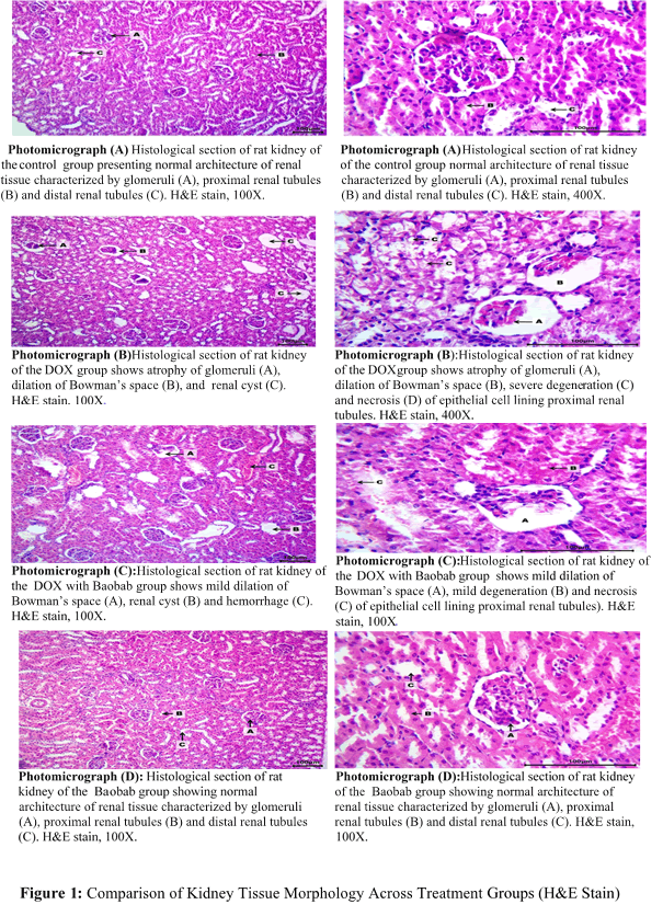
Descriptive histology result of kidney: Photomicrograph (A) The findings of the histological assessment of rat kidneys for the control group sections showed the normal architecture of renal tissue, Photomicrograph (B) DOX group may be indications of renal damage from DOX therapy in this group. One may note: Huge or atypical glomeruli, Tubule vacuolation, or gaps filled with fluid, lossof the proximal convoluted tubules’ brush border inflammatory cells penetrating. Photomicrograph (C) showed possibly demonstrate baobab’s ability to shield against harm brought on by DOX. In contrast to the DOX group, one may observe: Less drastic changes to the glomerular architecture. decreased vacuolation or elimination of the brush border. Photomicrograph (D) Treatment with baobab resemble the control group since normal kidney morphology is not anticipated to be substantially affected by Baobab alone (FIGURE 1).
FIGURE 1. Comparison of kidney tissue morphology across treatment groups (H and E stain).
Discussion
Descriptive histology result of liver
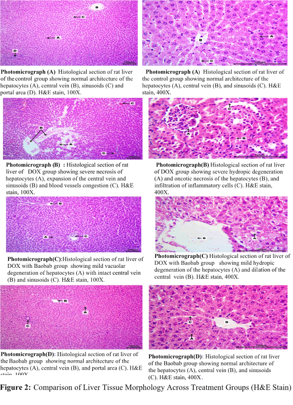
In control group showed normal liver architecture while in DOX group demonstrations severe impairment to the liver tissue caused by DOX treatment appeared Necrosis of hepatocytes with expansion of the sinusoids and central vein. This indicates blood vessel obstruction or congestion. DOX with baobab group showed improvement in liver morphology compared to the DOX group, suggesting a potential protecting of baobab some damage is still obvious degeneration of hepatocytes that denotes to swollen cells with pale, fluid-filled cytoplasm. Dilation of the central vein with some congestion is nonetheless current. Baobab alone does not appear to significantly influence healthy liver tissue (FIGURE 2).
FIGURE 2. Comparison of liver tissue morphology across treatment groups (H and E stain).
Rats and humans have very high genetic similarities (around 90%). This implies that it is often possible to induce disease and give medications, and similar biological processes in rats. Rats reproduce fast and are generally inexpensive and simple to grow in a scientific environment. Although rats are bigger than mice, which are also often employed in research, they are still manageable in terms of handling and housing. Additionally, their behavior is complicated enough to be useful in research on memory, learning, and other cognitive processes.
Current study demonstrated the dual role of doxorubicin as both a therapeutic agent and a potential source of organ toxicity. On the other hand, baobab plants, specifically Adansonia digitata show promise in protecting against hepato-renal damage induced by toxic agents.
The histological results of this study have shown that DOX treatment leads to substantial hepatic tissue damage, characterized by severe hydropic degeneration and oncotic necrosis of hepatocytes, dilation of sinusoids, and blood vessel congestion. These findings were consistent with the known hepatotoxic effects of DOX, which include oxidative stress and metabolic disruptions leading to cellular damage and inflammation. Also agree we have also demonstrated that DOX can cause degeneration, necrosis, and hyperplasia of Kupffer cells, as well as dilation of the central vein and sinusoidal congestion. They found the infiltration of inflammatory cells is a common response to DOX-induced liver injury, further exacerbating tissue damage and contributing to the overall pathology.
The results of this study consistent with Kewedar, 2023 found the known nephrotoxic effects of doxorubicin, which have been documented in various studies. As an illustration, it has been shown that doxorubicin therapy in rats results in a considerable necrosis of the renal tubules and glomeruli, as well as interstitial cell infiltration and a reduction in glomerular diameter. Furthermore, originate vacuolar degeneration and necrosis of the epithelial cells covering the renal tubules are consistent with observations made in studies that showed doxorubicin produced substantial histological changes, such as expanding of the tubular cells, loss of the brush boundary, and unclear degeneration. As well as they enlargement of Bowman’s space and improved collagen content experimental in doxorubicin-preserved kidneys further substantiate these findings, representing uncompromising structural impairment. Also, the admission of eosinophilic proteinaceous material and the attendance of renal cysts are suggestive of chronic kidney damage, which has been experiential in models of doxorubicin-induced nephrotoxicity.
The results of the present study presented the Baobab treatment led to minimal renal hypertrophy and preserved the reliability of the glomerular and tubular structures, representing a potential improvement of glomerulosclerosis and tubular atrophy in rats with infravesical blockade. Furthermore, we found the multidirectional action of neurotrophic factors present in Baobab, which contributed to the partial restoration of the morphological structure of rat kidneys affected by the experimental condition.
Also agree with that we found that the treatment with Adansonia digitata fruit pulp led to a dose-dependent partial reduction in lesion intensity in the liver tissue, while the heart and kidney tissues mostly returned to normal histology, indicating healing effects. And the inflammatory infiltration in the tissues was eliminated, showing that the fruit pulp had strong anti-inflammatory properties, especially at higher doses. As well as we found rats treated with both lead acetate and Adansonia digitata showed less liver damage, with only mild fat accumulation and some large fat droplets in liver cells. Rats treated with both lead acetate and Adansonia digitata showed normal kidney structures with only minor damage to kidney tubules. Additionally we found treatment with flavonoids fractions of Adansonia digitata (25, 50, and 75 mg/kg) resulted in mild liver steatosis and some microvesicular fatty droplets, indicating partial protection against HgCl2 induced damage. And the kidneys of rats treated with Flavonoids fractions of Adansonia digitata showed less degeneration compared to those treated only with HgCl2, suggesting a protective effect of FAD on renal tissues and consistent with found that at high doses (800 mg/kg), the Adansonia digitata leaf extract caused slight damage to the liver and kidneys, including glomerular and hepatic necrosis, and increased inflammatory cells in the spleen tissue.
Conclusion
The protective effects of using natural product like Adansonia digitata are likely due to its antioxidant and anti-inflammatory properties, which help restore the normal structure of liver and kidney tissues and prevent damage caused by free radicals produced by doxorubicin.
Acknowledgement
Our appreciation to everyone who assisted us to successfully complete this research. First pharmacy collage Nineveh university for support and help to complete the study. We also appreciate the technical support and provision of the facilities and resources required for carrying out the histological studies by the laboratory personnel at veterinary college/University of Mosul.
References
- Johnson-arbor K, Dubey R. Doxorubicin. (2020).
[Google Scholar] [PubMed]
- Zhao H, Yu J, Zhang R, et al. Doxorubicin prodrug-based nanomedicines for the treatment of cancer. Eur J Med Chem. 5, 115612 (2023).
[Crossref] [Google Scholar] [PubMed]
- Li X. Doxorubicin-mediated cardiac dysfunction: Revisiting molecular interactions, pharmacological compounds and (nano) theranostic platforms. Environ Res. 24, 116504 (2023).
[Crossref] [Google Scholar] [PubMed]
- Akin AT, Öztürk E, Kaymak E, et al. Therapeutic effects of thymoquinone in doxorubicinâinduced hepatotoxicity via oxidative stress, inflammation and apoptosis. Anat Histol Embryol. 50, 908-917 (2021).
[Crossref] [Google Scholar] [PubMed]
- Hsieh PL, Chu PM, Cheng HC, et al. Dapagliflozin mitigates doxorubicin-caused myocardium damage by regulating AKT-mediated oxidative stress, cardiac remodeling, and inflammation. Int J Mol Sci. 23, 10146 (2022).
[Crossref] [Google Scholar] [PubMed]
- Luo T, Yang S, Zhao T, et al. Hepatocyte DDX3X protects against drug-induced acute liver injury via controlling stress granule formation and oxidative stress. Cell Death Dis. 14, 400 (2023).
[Crossref] [Google Scholar] [PubMed]
- Ali AA, Saad EB, ElâRhman RH, et al. Impact of peroxisome proliferator activated receptor agonist drugs in a model of nephrotoxicity in rats. J Biochem Mol Toxicol. 37, e23350 (2023).
[Crossref] [Google Scholar] [PubMed]
- Kour H, Singh A, Jaiswal P, et al. Screening models of nephrotoxicity and their molecular mechanism. World J Bio Phar Health Sci. 13, 234-51(2023).
- On JY, Kim JM, Kothari D, et al. Antioxidant Properties and Kidney Cell Protection by the Extracts of Curcuma longa, Artemisia princeps, Salicornia herbacea, and Schisandra chinesis. Fermentation. 8, 702 (2022).
- Bag A, Byahut A, Khandelwal B. Medicinal plants with kidney-protecting effect in diabetic nephropathy. Curr Sci. 123(2022).
- Akintayo ET. Chemical Composition, Cacium, Zinc and Phytate Interrelationships in Baobab (Adansonia digitata) Seed Flour.
- Ofori H, Addo A. A review of baobab (Adansonia digitata) fruit processing as a catalyst for enhancing wealth and food security. J Ghana Instit Eng. 23, 34-43 (2023).
- Silva ML, Rita K, Bernardo MA, et al. Adansonia digitata L.(Baobab) bioactive compounds, biological activities, and the potential effect on glycemia: a narrative review. Nutrients. 15, 2170 (2023).
[Crossref] [Google Scholar] [PubMed]
- Goel C, Dutta R. Antimicrobial Activity of Leaf and Stem Extracts of Adansonia digitata. 24, 104-13 (2023).
- Komane B, Kamatou G, Mulaudzi N, et al. Adansonia digitata. 1-39(2023).
- Bashir KU, Tijjani H. Glucose Lowering Effects and in vitro α-Amylase and α-Glucosidase Inhibitory Potential from Aqueous Extract of Adansonia digitata (Baobab) Seed. Med Sci Forum. 14, 66 (2022).
- Uhuo EN, Egba SI, Nwuke PC, et al. Antioxidative properties of Adansonia digitata L. (baobab) leaf extract exert protective effect on doxorubicin-induced cardiac toxicity in Wistar rats. Clin Nutr Open Sci. 45, 3-16 (2022).
[Crossref] [Google Scholar] [PubMed]
- Egbadzor KF, Akuaku J. Prospects of raising baobab (Adansonia digitata L.) to fruiting in two years. Tree Fores People. 8, 100232(2022).
- Adesina JA, Zhu J. A review of the geographical distribution, indigenous benefits and conservation of african baobab (Adansonia digitata L.) tree in sub-saharan Africa.
- Ibrahim AM, Yassin KE. A Note on the Effect of Storage on Physicochemical Properties of Baobab (Adansonia digitata L.). 30, 95-95(2022).


