Case Report - Imaging in Medicine (2022) Volume 14, Issue 6
A Rare Case of Synchronous Gastric Signet Ring Cell Adenocarcinoma Metastasis at the Right Colon Angle
SeifeddineBaccouche1, Sarraj Achref1, Mohamed Hajri1, Osman Rania1, Wael Ferjaoui1*, Ahlem Lahmer2, Sana ben Slama2, DhouhaBacha2
1Department of General surgery, Mongi Slim University Hospital, Faculty of medicine of Tunis, University of Tunis el Manar
2Department of pathology, Mongi Slim University Hospital,Faculty of medicine of Tunis, University of Tunis el Manar
- *Corresponding Author:
- Wael
Ferjaoui
Department of General surgery, Mongi Slim University Hospital, Faculty of medicine of Tunis, University of Tunis el Manar
E-mail: farjaouiwael4@gmail.com
Abstract
A Rare Case of Synchronous Gastric Signet Ring Cell Adenocarcinoma Metastasis at the Right Colon Angle Although it can affect any part of the gastrointestinal system, signet ring cell carcinoma (SRC) is a common malignant entity of stomach cancers.
We discuss the case of a patient who had a prior history of gastric adenocarcinoma with signet ring cells, for which he underwent a partial gastrectomy with a gastro-jejunal anastomosis.
Four months after surgery, he presented an acute postoperative blockage. Operative findings and pathology exams concluded the presence of SRC at the right angle of the colon.
In patients with obstructive symptoms after surgery for SRC neoplasms of the stomach, the possibility of a second lesion should be considered among the differential diagnoses.
Keywords
Signet ring cell carcinoma • Stomach • Gastrointestinal system • Surgery
Background
Globally, gastric cancer is the third highest cause of cancer death [1]. Signet ring cell carcinoma (SRC) is widely known among the various gastric cancers for having a rapid progression and thus a bad prognosis.
Although it can affect any part of the gastrointestinal tract, it is a typical malignant entity of gastric cancers.
SRC is described as a stomach adenocarcinoma with more than a 50% contingency of solitary cells or small groups of cells distributed in a fibrous stroma including intra-cytoplasmic mucin (according to the WHO classification). The nucleus may be pushed to the periphery by this mucin, resulting in a chastened ring cell appearance [2].
Atypical locations of primary and metastatic SRC include the small intestine and colon.
In the present report we demonstrate a case of metastatic gastric SRC of the colon.
Clinical Case
A 53-year-old Patient was diagnosed withundifferentiatedantral adenocarcinoma that was detected during an oeso-gatro-duodenal fibroscopy.
The patient underwent surgery.
There were no local invasion nordistant metastases. According to Finsterer, a partial gastrectomy was performed with a gastro-jejunal anastomosis. The immediate postoperative recovery was straightforward. The anatomopathological analysis concluded a poorly differentiated adenocarcinoma with a catkin-ring independent cell contingent and lymph node invasion.
Four month later, the patient went to the emergency room with bowls obstruction that had been developing for 72 hours, along with occasional episodes of vomiting.
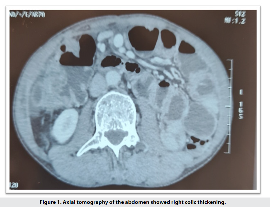
The patient appeared to be in good general health. Clinical exam demonstrated a minor abdominal meteorism and widespread tympany without peritoneal symptoms. The scar of the prior laparotomy was solid. No lump was discovered during the rectal examination. An abdominal CT scan with contrast injection revealed ascending colon distension upstream of a thickening of the right colonic angle (Figure 1) and an incontinent ileo -caecal valve with no symptoms of digestive distress.
The patient has had a nasogastric tube but the bowls obstruction persisted. We decided to operate.
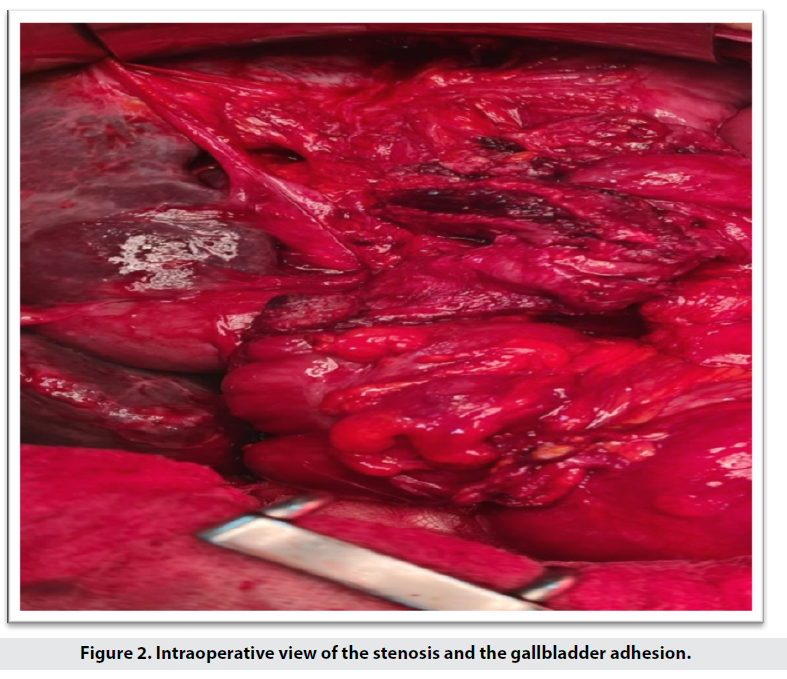
We discovered a thickening at the level of the right colonic angle that had a solid consistency, as well as intimate adhesions with the gallbladder (Figure 2).
Figure 2: Intraoperative view of the stenosis and the gallbladder adhesion.
The gallbladder was removed in one piece after a right hemicolectomy with full resection of the mesocolon. An ileo-colonic anastomosis was used to reestablish digest if continuity.
Anatomopathological exam determined to be a Signet ring cell carcinoma infiltrating the gallbladder.
The second day after surgery, the patient died abruptly. A massive embolism of the pulmonary artery is the probable cause of death.
Discussion
The third biggest cause of cancer death is gastriccancer [1].
Hematogenous and lymphoid metastasis is known routes of gastric cancer metastasis.The most common type of metastasis ishematogenous. The liver, lung, and pancreas are the most common sites of gastric cancer metastases [3].
Gastric cancer is the most common primer lesion, followed by breast, kidney, prostate, and ovarian cancers. Metastases to the colon are extremely rare [4]. In the current case, the patient was diagnosed with gastric cancer metastatic to the right colon.
It is crucial to identify the three main pathways of gastric SRC adenocarcinoma dissemination: the gastrointestinal system, the circulatory system (hematogenous) and the lymphatic system (lymphogenic). Multiple lesions are frequently identified in the pattern of metastasis via the gastrointestinal lumen. The polypoid form of these metastases was common. Hematogenousmetastases, which form on the interior of the colonic wall and resemble a submucosal tumor from the lumen, have been described. The lesion has a morphology that is comparable to the one described during primary colorectal cancer in lymphatic metastases [3].
It is obvious from the above that when cancers of the same histologic type are discovered in both the stomach and the intestine, establishing whether the tumors constitute a primary or a dual tumor site is challenging.
When a poorly differentiated adenocarcinoma or signet cell carcinoma is discovered in both the stomach and the large intestine, the risk of metastasis from the stomach to the large intestine should be considered.
Browsing through literature, we have discovered a wide range of macroscopic manifestations of metastatic intestinal lesions. Polyps, ulcerations, and depressed lesions were the most common [1]. However, there is a paucity of information regarding gastric SRC metastatic lesions presenting as colonic stenosis.
The location of the synchronous metastases is another unique characteristic of our case. Sonoda et al. found 11 cases of primary gastric SRC with metastasis to the large bowel, with six cases having metastasis to the sigmoid colon and only one case having metastasis to the rectum in their assessment of the literature [1]. Our patient had an unusual right colon metastasis.
Conclusion
Gastric SRC metastasis to the colon as a stenotic development is a rare occurrence.
According to research, the majority of gastric SRC metastasis to the colon manifests as ulcerations, depressed or flat lesions.
Given the rarity of gastric SRC metastasis to the colon with stenosis formation, this case offers a chance to raise physician awareness and understanding about the search for possible colon metastasis during gastric.
References
- Mehershahi S, Mantri N, Sun H et al. Metastasis From Gastric Signet Ring Cell Adenocarcinoma Presenting as a Rectosigmoid Stricture: A Rare Case. Cureus [Internet]. 12 (2020)
- Bosmon FT, Carneiro F, Hruban RH et al. WHO Classification of Tumours of the Digestive System [Internet]. World Health Organization. (2010).
- Sonoda H, Kawai K, Yamaguchi H et al. Lymphogenous metastasis to the transverse colon that originated from signet-ring cell gastric cancer: A case report and review of the literature. Clin Res Hepatol Gastroenterol. 41, e81–6 (2017).
- Ogiwara H, Konno H, Kitayama Y, et al. Metastases from gastricadenocarcinoma presenting as multiple colonic polyps: report of a case. Surg today. 24, 473-475 (1994).
Indexed at, Google Scholar, Crossref
Indexed at, Google Scholar, Crossref


