News and Views - Imaging in Medicine (2013) Volume 5, Issue 2
Seven signs could help rule out pediatric CT in traumatic abdominal injuries CT texture analysis could predict prognosis in esophageal cancer, New technology developed to improve reviewing images during surgical procedures, White matter hyperintensities volume associated with Alzheimer's disease
Sarah Miller*Commissioning Editor, Imaging in Medicine
- Corresponding Author:
- Sarah Miller
Commissioning Editor
Imaging in Medicine
E-mail: s.miller@futuremedicine.com
Abstract
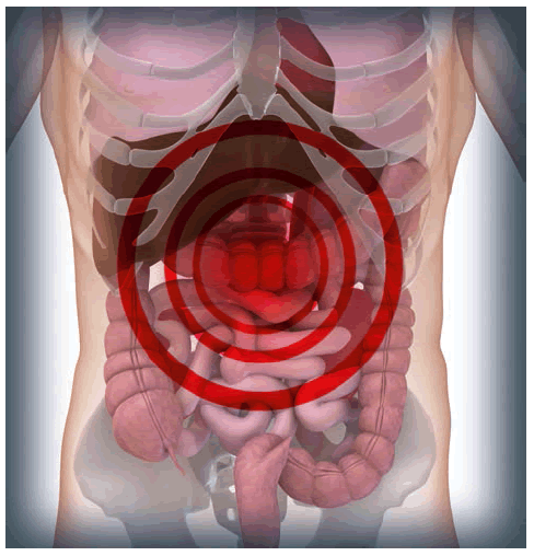
Seven signs could help rule out pediatric CT in traumatic abdominal injuries
A recently published prospective study from the Pediatric Emergency Care Applied Research Network (PECARN) in the USA has suggested a prediction rule to identify children who may not need a CT scan following blunt abdominal trauma. Those identified as very low risk of clinically important blunt abdominal injuries could be spared the significant radiation risk associated with pediatric CT.
“CT scans involve significant radiation risk, especially for children, who are more vulnerable than adults to radiation’s effects,” explained principal investigator and lead author of the study James Holmes, Professor of Emergency Medicine at the UC Davis School of Medicine (CA, USA). “We have now identified a population of pediatric patients that does not typically benefit from a CT scan, which is an important step in reducing radiation exposure.”
The recent study, published online ahead of print in Annals of Emergency Medicine, prospectively enrolled 12,044: children (median age 11.1 years) with blunt torso trauma from the 20 emergency departments involved in PECARN. Patients sustained their injuries from, for example, a fall, assault or car or bike crash and were being considered for acute intervention (e.g., therapeutic laparotomy, angiographic embolization, blood transfusion for abdominal hemorrhage and so on) in the emergency department.
Instead of performing a CT, the PECARN investigators only considered historical and physical examinations in study participants. They identified a sevensign prediction rule for identifying those who were at a very low risk of clinically important blunt abdominal injuries, for whom a CT would probably not provide any additional useful information for clinical decisions.
In order of importance, no evidence of abdominal wall trauma or seat belt sign, Glasgow Coma Score >13 and a lack of abdominal tenderness were identified as three important factors in predicting whether or not a CT would be necessary. These were followed by no evidence of thoracic wall trauma, no complaints of abdominal pain, no decreased breath sounds and an absence of vomiting.
The researchers reported that children who met all seven criteria had only a 0.1% chance of a clinically important injury requiring acute intervention, and in these patients a CT would be unlikely to reveal anything else. Authors propose that the risk of developing a future cancer from the radiation of a CT scan in this situation would be greater than the risk of the child developing a significant medical problem from the injury. However, they caution that clinical judgment remains important in deciding whether to perform a CT in each case.
While previous PECARN studies have already led to new standards of care for pediatric patients with head trauma, diabetic crisis and infections, these most recent findings relating to abdominal blunt trauma CT require external validation before implementation.
A further PECARN study is currently investigating the use of ultrasonography in pediatric cases of blunt abdominal trauma; another potential means of reducing radiation exposure in this patient group.
– Written by Sarah Miller
Sources: Holmes JF, Lillis K, Monroe D et al. Identifying children at very low risk of clinically important blunt abdominal injuries. Ann. Emerg. Med. doi:10.1016/j.annemergmed.2012.11.009 (2013) (Epub ahead of print); UC Davis Health System. Needless abdominal CT scans can be avoided in children, study says: www.ucdmc. ucdavis.edu/publish/news/newsroom/7428
CT texture analysis could predict prognosis in esophageal cancer
A team of UK-based researchers has recently presented findings which suggest that assessing intratumoral heterogeneity using postprocessing CT texture analysis could provide prognostic biomarkers for primary esophageal cancer patients receiving neoadjuvant chemotherapy. There are currently no established imaging or histological biomarkers for identifying good responders or those with a good prognosis who could benefit from surgery. The study abstract was presented at the 2013 Cancer Imaging and Radiation Therapy Symposium (FL, USA) in February 2013.
Curative surgery has reported poor outcomes in early stage esophageal cancer and neoadjuvant therapy is given to promote local and distant tumor control. This recent study examined 31 esophageal cancer patients who received platinum- and fluorouracil-based chemotherapy followed by surgery. Patients were identified retrospectively from institutional databases.
Postprocessing texture analysis was performed on the patients’ staging and postchemotherapy CT scans using proprietary software. Texture parameters examined were mean-gray level intensity, entropy, uniformity, kurtosis, skewness and standard deviation of histogram. These were derived for four filter values in order to highlight structures of different spatial width (1.0 [fine texture], 1.5–2.0 [medium] and 2.5 [coarse]).
Several baseline and post-treatment texture parameters were identified as significant positive prognostic factors in this patient group. For example, a smaller change in skewness following chemotherapy was a significant prognostic factor. In addition, baseline and post-treatment standard deviation were found to have significant associations with pathological tumor response.
“Although these results are for a very small number of patients, they suggest that the tumoral texture features may provide valuable information that could help us to distinguish which patients will do well following chemotherapy and which ones will do poorly,” explained lead author Connie Yip (King’s College London [UK] and National Cancer Centre [Singapore]). “As a biomarker for treatment efficacy, this technique could save patients from unnecessary surgery and provide more definitive guidance in developing patient treatment plans with improved outcomes,” she concluded.
– Written by Sarah Miller
Source: ASTRO. CT texture analysis of tumors may be a valuable biomarker in localized esophageal cancer: www.astro.org/News-and- Media/News-Releases/2013/CT-texture-analysisof- tumors-may-be-a-valuable-biomarker-inlocalized- esophageal-cancer.aspx
New technology developed to improve reviewing images during surgical procedures
Engineers from Purdue University (IN, USA) have described their attempts to improve upon one of the most common pieces of equipment in surgical theatres, the computer workstation. In a development that the researchers hope can improve surgical procedure times and infection rates, the Purdue team have described a gesture-based control scheme for surgical computing equipment.
Describing the opportunity for improvement, Juan Pablo Wachs from Purdue University said, “Computers and their peripherals are difficult to sterilize, and keyboards and mice have been found to be a source of contamination. Also, when nurses or assistants operate the keyboard for the surgeon, the process of conveying information accurately has proven cumbersome and inefficient since spoken dialogue can be time-consuming and leads to frustration and delays in the surgery.”
The researchers developed a system that utilized depth-sensing cameras and specialized algorithms to allow a program to recognize ten hand gestures to provide a variety of manipulation options for a surgeon. The results of the study are outlined in the team’s article published in the Journal of the American Medical Informatics Association.
“A major challenge is to endow computers with the ability to understand the context in which gestures are made and to discriminate between intended gestures versus unintended gestures,” Wachs said. In their study, an image navigation and manipulation task was performed to assess the effectiveness of the system; by including contextual information in the set up, the team was able to reduce the rate of false-positive gesture identification from 20.8 to 2.3%.
Based on their findings, the Purdue team believes that in the future, systems such as this could replace conventional mouse and keyboard systems in the operating room to improve patient care during surgery.
– Written by Sean Fitzpatrick
Sources: Jacob MG, Wachs JP, Packer RA. Handgesture- based sterile interface for the operating room using contextual cues for the navigation of radiological images. J. Am. Med. Inform. Assoc. doi:10.1136/amiajnl-2012–001212 (2012) (Epub ahead of print); Purdue University. Surgeons may use hand gestures to manipulate MRI images in OR: www.purdue.edu/newsroom/ releases/2013/Q1/surgeons-may-use-handgestures- to-manipulate-mri-images-in-or.html
White matter hyperintensities volume associated with Alzheimer’s disease
Research published recently in JAMA Neurology suggests that small-vessel cerebrovascular disease, which involves the interruption of blood circulation within the brain, may be a predictive factor of Alzheimer’s disease (AD). Led by scientists from Columbia University (NY, USA), the team’s research indicates that small-vessel cerebrovascular disease may be implicated alongside b -amyloid accumulation in the development of the disease.
To obtain their findings, the investigators used data from the Alzheimer’s Disease Neuroimaging Initiative database, taking the results of Pittsburgh compound B (PiB)-PET and structural MRI scans from 20 individuals diagnosed with AD, 59 diagnosed with mild cognitive impairment and 21 cognitively healthy control subjects. The team examined PiB positivity and the volume of white matter hyperintensities (WMH), a symptom associated with small-vessel cerebrovascular disease, in each image. The data from these scans were then analyzed to determine whether there was a correlation between PiB status, WMH volume and the cognitive health of the scanned individuals.
The team found that PiB positivity and increased total WMH volume were both independently associated with AD. PiB-positive individuals diagnosed with AD were shown to have greater WMH volume than cognitively healthy control subjects. Interestingly, both WMH volume and PiB status among the individuals with mild cognitive impairment were effective as predictors of a future diagnosis of AD, suggesting that the team’s findings could be of significant value to clinicians.
Speaking to Future Medicine about the results of their work, lead researcher Adam Brickman (Columbia University, NY, USA) informed them that their findings add “to a growing body of literature that highlights the importance of cerebrovascular disease in the clinical presentation, and possibly the pathogenesis, of AD.”
As the “risk factors for cerebrovascular disease, which include conditions such as hypertension, diabetes and obesity, are largely preventable through lifestyle modification” and are “treatable pharmacologically,” Brickman believes their research “suggests that lifelong management of vascular risk factors may delay the onset, mitigate or even prevent AD.”
When asked about his plans for future research, Brickman indicated that work in this area is ongoing. His team “are beginning to conduct studies to try to understand whether there is a mechanistic relationship between the lesions we see on MRI scans and primary AD pathology.” They are using various techniques to draw their findings, “including conducting MRI scans on post-mortem tissue and using histological techniques to study radiologically normal and abnormal tissue.”
– Written by Michael Mansbridge
Sources: Provenzano F, Muraskin J, Tosto G et al. White matter hyperintensities and cerebral amyloidosis: necessary and sufficient for clinical expression of Alzheimer disease? JAMA Neurol. doi:10.1001/jamaneurol.2013.1321 (2013) (Epub ahead of print); Medical News Today. Brain lesions can predict Alzheimer’s diagnosis: www. medicalnewstoday.com/articles/256587.php


