News and Views - Imaging in Medicine (2011) Volume 3, Issue 4
News & Views in ... Imaging in Medicine
Abstract
Brain imaging study looks into the differences in children with ADHD
New findings, published in The Clinical Neuropsychologist, have begun to uncover some of the differences in brain activity and development in children with ADHD. The preliminary study in question utilized magnetic resonance imaging to view differences in brain structure in 26 preschool children (13 with ADHD and 13 normal controls), between the ages of 4–5. The children selected were diagnosed using the standard DSM IV criteria.
“...the researchers were also able to demonstrate that the size of the nucleus in these individuals correlated with parental ratings for their respective children, of hyperactivity and impulsive symptoms.”
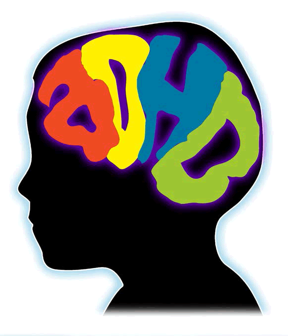
The results demonstrated that children with ADHD had a signif icantly smaller sized caudate nucleus. Typically this region of the brain has been associated with threshold control and learning.
When speaking to Future Medicine, Mark Mahone, first author of the paper and a researcher at the Kennedy Krieger Ins t itute (MD, USA) said, “By the age of 4 years, as many as 40% of children have sufficient problems with attention to be of concern to parents and preschool teachers, and ADHD has become the most commonly diagnosed form of psychopathology in the preschool years.
Since ADHD is a disorder that by definition has its onset prior to age 7 years, it is critical to examine children prior to that age to better understand the neurobiological course of the disorder. However, virtually all of the neuroimaging studies of children with ADHD have included only children of school age (i.e., age 6 years and older).”
Whilst other studies have looked at the metabolism of the caudate nucleus and ADHD, this study has focused on the structural component of the caudate nucleus. In addition to having a reduced volume, the researchers were also able to demonstrate that the size of the nucleus in these individuals correlated with parental ratings for their respective children, of hyperactivity and impulsive symptoms.
“The findings from our study revealed early anomalies (reduced volumes) in the development of the caudate nucleus among preschool children presenting with symptoms of ADHD, and a strong association between bilateral caudate volumes and severity of hyperactive/impulsive (but not inattentive) symptoms,” explained Mahone.
“With ADHD being the most commonly diagnosed behavioral problem in children, it is important to continue research into the fundamentals of the disease.”
When asked about the future impact of the work, Mahone replied: “The present findings highlight insights gained by examining early brain development among children with ADHD, and how findings among younger children may differ from those observed in older children. We seek to make more targeted behavioral and pharmacological interventions earlier in children with ADHD, in order to improve developmental and functional outcomes in this group.”
Mahone stresses that these preliminary data are part of an ongoing study to further understand the link between development and ADHD: “These findings are part of a longitudinal study of preschool children with ADHD. We plan to follow 100 preschool children (50 at risk for ADHD, 50 control) in order to uncover a critical link between brain and behavioral development in ADHD, with the ultimate goal of identifying important early biomarkers for the disorder. In particular, this study should ultimately allow for the description and comparison of the early developmental changes in preschoolers that contribute to the behavioral phenotype, and ultimately the formal diagnosis of ADHD, as well as those factors (anatomy, cognition, behavior and environment) that are associated with ‘protection from’ the disorder.”
“We seek to make more targeted behavioral and pharmacological interventions earlier in children with ADHD, in order to improve developmental and functional outcomes in this group.”
With ADHD being the most commonly diagnosed behavioral problem in children, it is important to continue research into the fundamentals of the disease. When one considers how much of an impact ADHD can have upon a child’s normal development, it is clear that a greater understanding of the disorder is an important step in unraveling this complex problem.
Source: Mahone EM, Crocetti D, Ranta ME et al. A preliminary neuroimaging study of preschool children with ADHD. Clin. Neuropsychol. 1, 1–20 (2011).
Combined imaging agents may improve PET/CT imaging of cancer
A study recently published in The Journal of Nuclear Medicine has indicated that the imaging agents 18F NaF and 18F FDG in a single PET/CT scan may improve cancer detection. The research was presented at the SNM 58th Annual Meeting in San Antonio (TX, USA).
“What we stand to gain from combining these two agents is the diversification of tumor targeting.”
The multicenter study used 78 patients with proven cancers. To evaluate malignancy, all participants had separate PET/ CT scans with either 18F or 18F FDG as imaging agents, and also had combined PET/CT scans with both agents combined. Once central site then performed direct comparisons of the results.
The results showed combined 18F NaF and 18F FDG PET/CT to provide accurate interpretation of malignancies. In addition, the method of combining imaging agents allows the same information to be obtained from just one scanning session, which reduces patient exposure to radiation. For these reasons, the study authors believe PET/CT with both imaging agents to be a feasible method for cancer detection.
Andrei Iagaru, lead author of the study and assistant professor of radiology and nuclear medicine at Stanford University Medical Center, Stanford (CA, USA), explains, “During a time when healthcare costs are under intense scrutiny, consolidated procedures such as this one that provide comprehensive imaging data are a benefit to everyone – to clinicians, healthcare administrators and especially patients who would need only one scan instead of two.”
Iagaru adds, “What we stand to gain from combining these two agents is the diversification of tumor targeting. The beauty of it is that each agent has its own strength, and those are unified in the imaging. In combination they represent a powerful new tool for acquiring as much information as possible about the extent of a patient’s cancer.”
“...the study authors believe PET/CT with both imaging agents to be a feasible method for cancer detection.”
In order to identify the most suitable clinical scenarios for this method of imaging cancer, further evaluation of combined 18F NaF and 18F FDG in PET/CT is warranted.
Source: Iagaru A, Mittra E, Sathekge M et al. Combined 18F NaF and 18F FDG PET/CT: initial results of a multicenter trial. J. Nucl. Med. 52(Suppl. 1), 34 (2011).
Gamma imaging could provide superior cancer detection to ultrasound in dense breasts
Research performed at Hampton University (VA, USA) has shown promising results for the use of gamma imaging compared with ultrasound for detecting breast cancer. The results were recently published in the Journal of Nuclear Medicine.
The use of low-amplitude x-rays, or mammography, is currently the gold standard imaging modality used to detect breast cancer. In the case of women with dense breast tissue, support methods such as gamma imaging (BSGI) or ultrasound are used to provide more accurate diagnosis.
Imaging using BSGI provides functional images of breast physiology and does not rely on breast structure to identify lesions, which is why it has proven a successful tool for imaging dense breasts. BSGI works by using gamma radiation to monitor the uptake of a radiotracer, Technetium Tc99m Sestamini, by cancer cells.
The study involved using ultrasound and BSGI to perform breast imaging, then using biopsy as the gold standard to confirm malignancy. Results from the study demonstrated BSGI to be less likely to give negative results for malignant lesions, and less likely to give positive results for benign lesions.
Douglas Kieper, professor and nuclear medicine research supervisor at Hampton University and author of the study says, “A lot of white shows up on the mammograms of women with radiodense breasts, and it becomes a lot like trying to find one cloud in a cloudy sky. This study tells us that BSGI improves our ability to detect breast cancer when combined with other breast imaging techniques. What we are really looking at is the impact that BSGI and ultrasound have on breast cancer patient management. Comprehensive breast imaging including BSGI could improve breast cancer detection and provide a better prognosis for breast cancer patients.”
Source: Kieper D. Breast-specific gamma imaging compared with ultrasound in the management of patients with BIRADS 0 mammograms. J. Nucl. Med. 52(Suppl. 1), 246 (2011).
PET may predict response to HIV-associated tuberculosis treatment
A study recently published in the Journal of Nuclear Medicine has indicated that PET could be a useful tool for predicting whether tuberculosis treatment is working in individuals with HIV. The work was carried out by researchers at the Department of Nuclear Medicine at the University of Pretoria, South Africa.
Immunosuppressed individuals with HIV are prone to coinfection with multidrug resistant tuberculosis, which poses a major health threat to these patients. Assessing whether tuberculostatics, or drugs that inhibit the growth of tuberculosis, are working can take more time than these patients may have if their condition worsens. The use of PET to predict outcomes would allow for early intervention with alternative treatments.
HIV patients that had recently been diagnosed with tuberculosis were used in this study. All patients underwent wholebody 18F FDG PET prior to receiving tuberculostatics. Patients were then given a typical combination therapy of isoniazid, rifampicin and ethambutol. After four months of treatment patients were scanned a second time, and the percentage change in maximum standardized uptake value (SUVmax) was measured, which assesses glucose metabolic activity.
“Assessing whether tuberculostatics, or drugs that inhibit the growth of tuberculosis, are working can take more time than these patients may have if their condition worsens.”
From the PET results, it was found that the SUVmax of involved lymph nodes, number of lymph node basins and C-reactive protein levels were all higher in patients that were not responding to their treatment. Furthermore, the researchers determined that a cutoff of five or more lymph node basins allowed for separation of patients responding to treatment and patients not responding. These findings imply that imaging markers could be a potential method for predicting patient responses to tuberculostatics.
Lead author of the study, Mike Sathekge, explained, “Early detection of drug resistance of tuberculosis allows the initiation of an appropriate treatment, which may significantly affect patient survival. Currently, more than twothirds of patients with multidrug resistant tuberculosis die.”
According to Sathekge “18F-FDG PET has the potential to become a valuable clinical adjunct to the already available genotypic and phenotypic tests in patients for whom such tests are not feasible, are inconclusive, or are too lengthy to be of clinical relevance”.
Source: Sathekge M, Maes A, Kgomo M, Stoltz A, Van de Wiele C. Use of 18F-FDG PET to predict response to first-line tuberculostatics in HIV-associated tuberculosis. J. Nucl. Med. 52(6), 880–885 (2011).
Whole-body molecular PET/MR system now in clinical use in parts of Europe and America
The Siemens Biograph mMR integrated PET and magnetic resonance system has recently been given FDA approval and received a CE mark for use in parts of Europe. Siemens receipt of approval for their system followed assessment that the product meets safety expectations, provides clinical benefit and meets environmental protection requirements.
The Biograph mMR is the first integrated whole body molecular system with simultaneous acquisition technology. The system combines the benefits of 3T MRI with PET in one scan. The MRI provides a morphologic and functional image of patients, whereas PET produces a functional image from cellular processes and metabolism.
One system is used in the department of radiology at the university hospital in Tuebingen, Germany. The clinical director of the Department of Diagnostic and Interventional Radiology at the hospital, Claus Claussen, expects the Biograph mMR to provide greater precision compared with individual MRI and PET exams, both in diagnostics and treatment planning due to acquiring the data simultaneously.
Claussen states, “Patients will benefit from this new technology, as simultaneous molecular MR will give us additional information for personalized therapy management. Furthermore, the very good image quality will support us in the early diagnosis and understanding of highly relevant diseases.”
“The Biograph mMR is the first integrated whole body molecular system with simultaneous acquisition technology.”
Siemens Healthcare hope that their new multimodality system will be particularly valuable in the identification of neurologic, oncologic and cardiac conditions and in supporting therapy planning.
Source: Siemens healthcare: www.siemens.com


