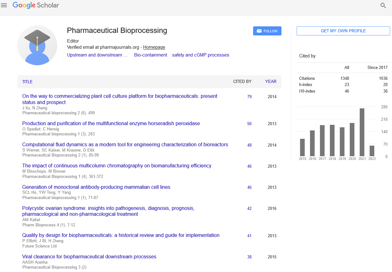
Mini Review - Pharmaceutical Bioprocessing (2023) Volume 11, Issue 1
Disulfide-rich recombinant proteins from Plasmodium falciparum can be efficiently produced using Lactococcus lactis as a platform.
David Gouveiac*
University of Cambridge, Cambridge, UK
University of Cambridge, Cambridge, UK
E-mail: gouveiacdavid@rediff.com
Received: 02-Jan-2023, Manuscript No. FMBP-23-86383; Editor assigned: 04-Jan-2023, PreQC No. FMBP-23-86383 (PQ); Reviewed: 18-Jan-2023, QC No FMBP-23- 86383; Revised: 23-Jan-2023, Manuscript No. FMBP-23-86383 (R); Published: 30-Jan-2023, DOI: 10.37532/2048-9145.2023.11(1).01-03
Abstract
Recently, it was discovered that Lactococcus lactis is an effective Gram-positive cell factory for the production of recombinant protein. This term “host” has been used by us and others to develop a few malaria vaccine candidates. Numerous clinical trials have confirmed this production system’s safety. In this study, we investigated L. lactis cell factories for the production of 31 representative Plasmodium falciparum antigens with varying sizes and predicted structural complexities. Among these antigens were eleven that had multiple predicted structural disulfide bonds, which are proteins that are thought to be difficult to produce.
Keywords
Plasmodium falciparum • Malaria • Disulfide-rich protein • Merozoite antigens • Lactococcus lactis
Introduction
The best expression system for making recombinant proteins is determined by a number of important factors, including yield, purity, and production cost. The biophysical and structural properties of the recombinant proteins cause expression yields to vary by orders of magnitude even if a single system is able to accommodate all of the proteins. The production of homogeneously folded disulfide-bonded proteins has been particularly challenging among the various proteins. Because it can export the recombinant protein into the culture supernatant, where it can be easily purified, the lactic acid bacteria Lactococcus lactis has gained importance as a host for heterologous protein expression. As a Gram positive bacterium, it does not produce endotoxins, and it is “generally recognized as safe.” Unwanted glycosylation of proteins has not been described. A low-cost production system is provided by the L. lactis expression system’s compatibility with large-scale upstream and downstream processes [1-4]. This expression system’s safety in humans has been demonstrated. We recently reported using L. lactis to successfully produce recombinant Pfs48/45, a disulfide-rich Plasmodium falciparum vaccine candidate that has been difficult to produce as a recombinant protein with correct conformation using a variety of other prokaryotic and eukaryotic recombinant protein expression systems. We have also shown that L. lactis can be used to express and purify 31 recombinant proteins from 25 different P. falciparum antigens, many of which contain multiple structural disulfide bonds, in this study. Recombinant antigens’ native structure and antigenic properties depend on the formation of the right disulfide bonds, as do the appropriate innate and adaptive immune responses. The Merozoite, P. falciparum’s transient extracellular stage of the asexual blood stage, is highly specialized for erythrocyte invasion, and the antigens chosen in this study are localized in various subcellular compartments. These include the proteins that make up the integral Merozoite membrane, the proteins that are associated with the peripheral surface, and proteins that are secreted from various secretory organelles like micromeres, rotaries, and dense granules. It is known that these proteins form functional complexes with the proteins that make up the Merozoite surface. Together, these merozoite surface protein complexes play a significant role in the invasion of erythrocytes. Several of these complexes have been identified as potential targets for malaria-protective immunity [5-8].
Purification of proteins
Batch fermentation and a two-step downstream purification procedure made up a simple workflow. The clones of L. lactis that expressed recombinant proteins were secreted into the culture medium, separated from the cellular biomass by centrifugation, and affinity purified with the His-tag using the His Trap HP-column. Ion-exchange chromatography was used to polish the recombinant protein-containing fractions following elution to further remove the contaminating host-cell protein (HCP). Recombinant cMSP33D7 serves as an illustration of how this method resulted in the production of a highly purified recombinant protein. Using the same method as before, recombinant R0- MSPDBL2 served as an example of recombinant R0-expressing proteins harvested from clarified culture medium. Prior to using an iron-exchange chromatography column to further purify the affinity-purified fusion protein, HCP, TEV, and the R0-fusion partner were separated from the target recombinant protein. Immunoblotting with antibodies against the His-tag and R0 was used to demonstrate that the carrier protein had been removed, and the identity and purity of the target protein were confirmed at each step.
Stabilizing structural domains with GLURP prevents thermal denaturation
On SDS-PAGE, its apparent molecular mass exceeds its predicted molecular mass as it migrates. Since boiling has been used to purify similarly disordered recombinant epsin 1 and AP180, we speculated that GLURP-R0 might stabilize its fusion partners against thermal denaturation because disordered proteins frequently resist heat. We cultured the MSPDBL2 construct without and with a R0-fusion partner in the initial experiment. More than 80% of the HCP was removed by centrifugation after the culture supernatants from the batch fermentation of recombinant clones expressing R0-fusion constructs were boiled and then immediately chilled in an ice-bath. While the R0-MSPDBL2 fusion protein remained in solution under these conditions, the MSPDBL2 recombinant protein was unable to withstand heat treatment. To separate TEV-protease, R0- fusion partner, and HCP from the target protein, the supernatant containing R0-MSPDBL2 was incubated with the TEV protease. The resulting mixture was then purified using an ironexchange chromatography column. SE-HPLC and ELISA were used to check the MSPDBL2- domain’s integrity. The analysis also included purified, unheated recombinant MSPDBL2 for comparison. According to SE-HPLC, the two MSPDBL2 recombinant proteins moved in the same direction with retention times of 5.54 minutes, indicating that heat treatment had no effect on protein stability or multimerization [9-10].
Discussion
Finding efficient and effective expression systems for the production of recombinant proteins is frequently complicated by the creation of new vaccines and sero-diagnostic tools. We have demonstrated that the L. lactis expression systems are ideal for the production of malaria antigens due to their ability to accommodate cysteine-rich proteins, their scalable fermentation process, their ability to secrete the recombinant protein into the culture medium, which makes the purification process easier, and their similar codon bias, which means that they do not require codon optimization prior to protein expression. E. wheat germ cell– free expression system and HEK293 cells have previously been utilized in the production of Plasmodium falciparum Merozoite antigens. Although each of these systems was successful with some antigens, it is difficult to produce the right post-translational modifications, which typically determine the quality and activity of the target recombinant protein, in each system. The endoplasmic reticulum (ER), which is an oxidizing environment, provides a setting for the formation of disulfide bonds in eukaryotic organisms like P. falciparum. The native disulfide bonds Jan be formed by eukaryotic expression systems. Prokaryotic organisms, on the other hand, lack the complex ER machinery and exhibit a wide range of mechanisms and capacities for the formation of protein disulfide bonds.
Conclusions
L. lactis recombinant proteins rich in disulfide can now be produced and purified using a novel method.
References
- Stirpe F. Ribosome-inactivating proteins.Toxicon.44, 371–383 (2004).
- Wang P, Tumer NE. Virus resistance mediated by ribosome inactivating proteins.Adv. Virus Res.55, 325–356 (2000).
- Olsnes S, Pihl A. Different biological properties of the two constituent peptide chains of ricin, a toxic protein inhibiting protein synthesis.Biochemistry.12, 3121–3126 (1973).
- Lord JM, Roberts LM, Robertus JD. Ricin: Structure, mode of action, and some current applications.FASEB J.8, 201–208(1994).
- Peumans WJ, Hao Q, Van Damme EJ. Ribosome-inactivating proteins from plants: More than N-glycosidases?FASEB J.15, 1493–1506 (2001).
- Stirpe F, Barbieri L. Ribosome-inactivating proteins up to date.FEBS Lett.195, 1–8 (1986).
- Kwon SY, An CS, Liu JR et al. Molecular cloning of a cDNA encoding ribosome-inactivating protein fromAmaranthus viridisand its expression inE. coli.Mol. Cells.10, 8–12 (2010).
- Lam YH, Wong YS, Wang B et al. Use of trichosanthin to reduce infection by turnip mosaic virus.Plant Sci.114, 111–117(1996).
- Lodge JK, Kaniewski WK, Tumer NE. Broad-spectrum virus resistance in transgenic plants expressing pokeweed antiviral protein.Proc. Natl. Acad. Sci. USA.90, 7089–7093 (1993).
- Carzaniga R, Sinclair L, Fordham-Skeleton AP et al. Cellular and subcellular distribution of saporins, type I ribosome-inactivating proteins, in soapwort.Plantae.194, 461–470(1994).
Indexed at, Google Scholar, Crossref
Indexed at, Google Scholar, Crossref
Indexed at, Google Scholar, Crossref
Indexed at, Google Scholar, Crossref
Indexed at, Google Scholar, Crossref
Indexed at, Google Scholar, Crossref
Indexed at, Google Scholar, Crossref