Case Report - Interventional Cardiology (2022)
Primary cardiac CD5-positive diffuse large B-cell lymphoma with bcl-6 gene rearrangement: A case report and review of literature
- Corresponding Author:
- Zhaoming Wang
Department of Pathology,
Zhejiang University School of Medicine First Affiliated Hospital,
Hangzhou,
China,
E-mail: wangzhaoming1963@163.com
Received date: 03-Oct-2022, Manuscript No. FMIC-22-61395; Editor assigned: 05-Oct-2022, PreQC No. FMIC-22-61395 (PQ); Reviewed date: 19-Oct-2022, QC No. FMIC-22-61395;Revised date: 26-Oct-2022, Manuscript No. FMIC-22-61395 (R);Published date: 02-Nov-2022, DOI: 10.37532/1755-5310.2022.14(S12).299
Abstract
Background: Primary cardiac CD5 positive Diffuse Large B-Cell Lymphoma (CD5+ DLBCL) with bcl-6 gene rearrangement is a rare subtype of DLBCL. The case is reported, along with a review of several characteristics of the disease.
Case presentation: We described a 64-year-old female patient who presented with a 12-month history of cough. Magnetic Resonance Imaging (MRI) revealed the mass in the right atrium and extending into the right ventricle.
Conclusion: CD5+ DLBCL was confirmed finally by pathological examination of the cardiac biopsy. The patient responded well to the modified R-CHOP regime. In a review of primary literatures, we know that factors contributing to the poor prognosis of DLBCL including CD5 positive, non-GCB immunophenotype, myc, bcl-2 and bcl- 6 gene rearrangements etc. Early diagnosis and aggressive treatment play a key role on improving the prognosis of primary cardiac CD5+ DLBCL.
Keywords
Cardiac • CD5 • Lymphoma • Histopathology • Gene rearrangement
Abbreviations
CD5+/- DLBCL: CD5 positive/negative Diffuse Large B-Cell Lymphoma; NHL: Non-Hodgkin’s Lymphoma; MRI: Magnetic Resonance Imaging; TEE: Transesophageal Echocardiography; ECG: Electrocardiogram; LDH: Lactate Dehydrogenase; FISH: Fluorescence In Situ Hybridization; CLL: Chronic Lymphocytic Leukemia; EBV: Epstein-Barr Virus; EBER: Epstein Barr Encoding Region; PET/CT: Positron Emission Tomography/Computed Tomography; FDG: Fluorodeoxyglucose; MCL: Mantle Cell Lymphoma; GCB: Germinal Center B-Cell; IHC: Immunohistochemistry; R-CHOP: Rituximab, Cyclophosphamide, doxorubicin, vincristine, and Prednisone; DA: Dose-Adjusted; EPOCH-R: Etoposide, Prednisone, vincristine, Cyclophosphamide, doxorubicin, and Rituximab; HD-MTX: High-Dose Methotrexate
Introduction
Diffuse Large B Cell Lymphoma (DLBCL)is the most common type of Non-Hodgkin’s Lymphoma (NHL), and representing 30%~40% of all NHLs with a high aggressiveness and poor prognosis, and occurring mostly in immune compromised patients. Primary cardiac CD5 positive Diffuse Large B-Cell Lymphoma (CD5+ DLBCL) is extremely rare subtype of DLBCL that involves only the heart and/or pericardium, accounting for <2% of all primary cardiac tumors [1,2]. Gene expression studies reveal that CD5+ DLBCL is distinct from other DLBCL. However, the diagnostic criteria and molecular features are still unclear. Herein, we report a patient who suffered from primary cardiac CD5 + DLBCL and summary its clinical characteristics by reviewing literatures.
Case Presentation
Clinical history
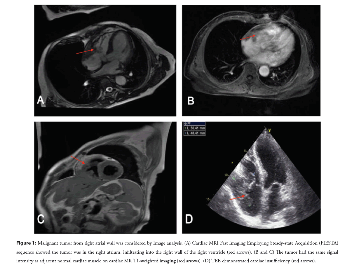
A 64-year-old female presented with a 12-month history of cough. No facial puffiness, exertional chest tightness, and dyspnoea, absent lower limb oedema and no lymphadenopathy. Magnetic Resonance Imaging (MRI) revealed a mass in the right atrium, infiltrating into the right wall of the right ventricle. MR imaging with dynamic enhancement showed a low density in the foci compared with the cardiac cavity (Figures 1A-1C). Transesophageal Echocardiography (TEE) demonstrated the mass induced severe cardiac insufficiency (Figure 1D). No space occupying lesions (no occupied space lesions) were found in the lung, liver, spleen and kidney. The patient had higher Lactate Dehydrogenase (LDH), creatine phosphokinase, hydroxybutyrate dehydrogenase in serum than healthy person.
Figure 1: Malignant tumor from right atrial wall was considered by Image analysis. (A) Cardiac MRI Fast Imaging Employing Steady-state Acquisition (FIESTA) sequence showed the tumor was in the right atrium, infiltrating into the right wall of the right ventricle (red arrows). (B and C) The tumor had the same signal intensity as adjacent normal cardiac muscle on cardiac MR T1-weighted imaging (red arrows). (D) TEE demonstrated cardiac insufficiency (red arrows).
Pathological findings
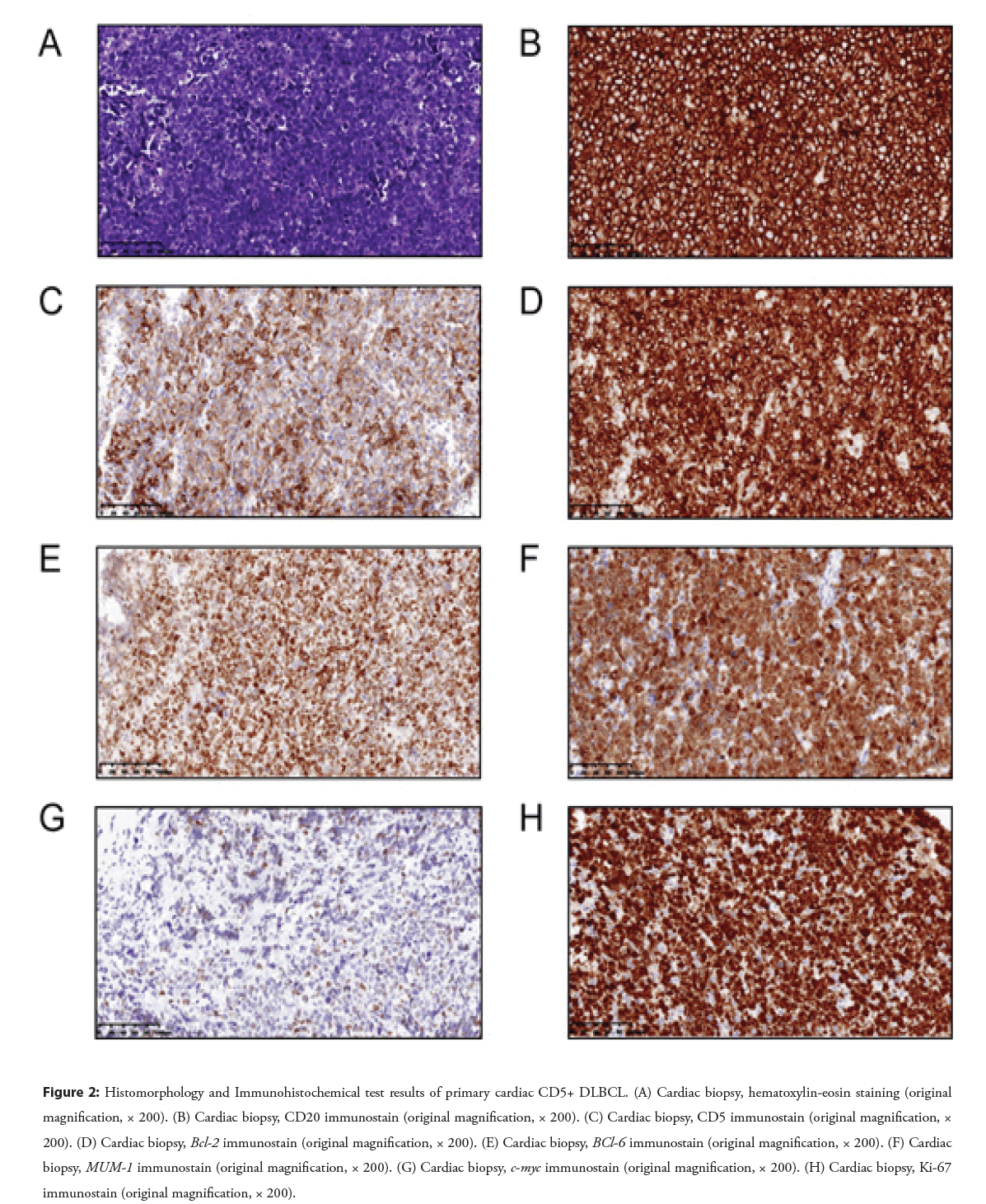
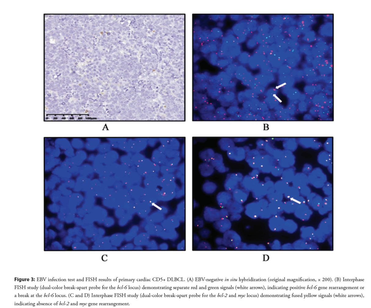
The macroscopy pathology showed that the total size of gray and broken tough tissue was about 0.5 × 0.5 × 0.2 cm. Histopathological examinations revealed the neoplastic proliferation of large lymphoid cells with round to irregular nuclear contours, vesicular nuclei, and single to multiple nucleoli (centroblastic and immunoblastic morphology) (Figure 2A). Immunohistochemical staining were performed and showed that the large neoplastic lymphoid cells were positive for CD20 (Figure 2B), CD5 (Figure 2C), Bcl-2 (Figure 2D), Bcl-6 (Figure 2E), MuM-1 (Figure 2F), Cmyc (Figure 2G) with a high Ki-67 proliferative rate (85%) (Figure 2H), while were negative for CD3, CD10, CyclinD1. No EBV infection was confirmed by Epstein Barr Encoding Region (EBER)-in situ hybridization (Figure 3A). Fluorescence In Situ Hybridization (FISH) studies were carried to detect myc, bcl-2, bcl-6 gene rearrangement and showed positive for bcl-6 gene rearrangement (Figure 3B) and negative for myc and bcl-2 gene rearrangement (Figures 3C and 3D). Thus, a final diagnosis of primary cardiac CD5-positive Diffuse Large B-Cell Lymphoma (CD5+ DLBCL) with bcl-6 rearrangement was made.
Figure 2: Histomorphology and Immunohistochemical test results of primary cardiac CD5+ DLBCL. (A) Cardiac biopsy, hematoxylin-eosin staining (original magnification, × 200). (B) Cardiac biopsy, CD20 immunostain (original magnification, × 200). (C) Cardiac biopsy, CD5 immunostain (original magnification, × 200). (D) Cardiac biopsy, Bcl-2 immunostain (original magnification, × 200). (E) Cardiac biopsy, BCl-6 immunostain (original magnification, × 200). (F) Cardiac biopsy, MUM-1 immunostain (original magnification, × 200). (G) Cardiac biopsy, c-myc immunostain (original magnification, × 200). (H) Cardiac biopsy, Ki-67 immunostain (original magnification, × 200).
Figure 3: EBV infection test and FISH results of primary cardiac CD5+ DLBCL. (A) EBV-negative in situ hybridization (original magnification, × 200). (B) Interphase FISH study (dual-color break-apart probe for the bcl-6 locus) demonstrating separate red and green signals (white arrows), indicating positive bcl-6 gene rearrangement or a break at the bcl-6 locus. (C and D) Interphase FISH study (dual-color break-apart probe for the bcl-2 and myc locus) demonstrating fused yellow signals (white arrows), indicating absence of bcl-2 and myc gene rearrangement.
After diagnosis, the patient was placed on a modified R-CHOP (Rituximab, Cyclophosphamide, Hydroxydaunorubicin (Adriamycin), Oncovin (vincristine), and Prednisone) chemotherapy regimen and her symptoms were improved. The patient is still alive after the initiation of therapy. PET/CT showed no obvious morphological changes in the right atrium, and FDG metabolism was normal in the right atrium and the whole body (including brain tissue). The further following-up is performing.
Results and Discussion
CD5 as a transmembrane receptor that mainly regulates T cell functions and development is also expressed by a small subset of normal B cells. It contributes to the progression of tumor by inhibiting the T cell response and protecting B cells from B Cell Receptor (BCR) signaling-mediated apoptosis [3]. Generally, CD5 is expressed in the B cell lymphoma such as Chronic Lymphocytic Leukemia (CLL) and Mantle Cell Lymphoma (MCL) [4]. In addition, a number of CD5 positive hairy cell leukemia cases have been reported [5]. Recent years, Several CD5+ DLBCL cases primary occurred in cardiac tissue, have been reported.
Xiao, et al. [6] reported a 64 years old female patient with primary CD5+ DLBCL in the right atrium. The patient’s main clinical manifestations were facial edema, tiring chest tightness and dyspnea, with jugular vein filling, and no edema of lower limbs. All the three reported cases by Soon, et al. [2] had right heart involvement. Only 1 case involved all 4 chambers of the heart. In our case, the mass occupied the right atrium, infiltrating into the right wall of the right ventricle. As we can see that, there were no specific clinical manifestations in the syndrome to help diagnosis. The most common symptoms are dyspnea, chest pain, and constitutional complaints such as fever, night sweats, loss of appetite, and weight loss. It usually involves the right heart, in particular the right atrium. Only less than 10% of cases show isolated left heart involvement.
Primary cardiac lymphomas account for only 1%-2% of all primary cardiac tumors [7,8]. Immunophenotypic subtyping of primary cardiac CD5+ DLBCL divided into cell-of-origin GCB and non-GCB subgroups is less commonly reported in the literature compared with DLBCL originating from other sites [9]. In our study, the case is belonged to non-GCB immunophenotype using the Hans IHC algorithm method, which express Bcl-6 and MuM-1, but negative for CD10. High-grade B-cell lymphomas with both c-myc and bcl-2 gene rearrangements or double-hit lymphomas are known to have an extremely poor prognosis [10,11]. The median survival time was less than 2 years [12]. In our case, bcl-6 gene rearrangement was detected positive. Primary studies have shown that there is no significant correlation between CD5 expression and myc, bcl-2 and/or bcl-6 gene rearrangement [4,13]. Further studies would be helpful to evaluate the prognostic relation of MYC, BCL-2 and BCL-6 protein co-expression or gene rearrangements with specific reference to primary cardiac CD5+ DLBCL.
There is still no standard and uniform scheme for the treatment of CD5+ DLBCL currently. Early diagnosis and aggressive treatment play a key role on improving the prognosis of primary cardiac CD5+ DLBCL. R-CHOP is still recommended in the current standard first-line therapy. In our case, R-CHOP chemotherapy is adopted and symptoms of the patient have been significantly relieved. However, CD5+ DLBCL occurs frequently in elderly patients who cannot tolerate high-dose chemotherapy and combination therapy. That will be a problem we must face in the future. A phase II study conducted by Miyazaki, et al. [14] showed that Dose-Adjusted (DA)-EPOCH-R (etoposide, prednisone, vincristine, cyclophosphamide, doxorubicin, and rituximab) combined with High-Dose Methotrexate (HD-MTX) might be a first-line therapy option for stage II-IV CD5+ DLBCL.
Conclusion
In conclusion, primary cardiac CD5+ DLBCL is an uncommon and aggressive subtype lymphoma. The factors promoting to the poor prognosis of DLBCL including CD5 positive, non-GCB immunophenotype, myc, bcl-2 and bcl-6 gene rearrangements or double/three-hit, Ki-67 positive rate more than 90% or a higher LDH value (more than 3 times compared with normal value). Earlier detection and diagnosis contribute to promote patient’s prognosis. Advanced radiological techniques and confirmatory histologic and/or cytologic diagnosis via periodic fluid drainage or variable endocardial biopsy techniques are the main diagnostic procedures.
Ethics Approval and Consent to Participate
The study was approved by the Human Ethics Committee of The First Affiliated Hospital Zhejiang University School of Medicine.
Consent for Publication
Written informed consent for publication of patient clinical details and/or clinical images was obtained from the patient. A copy of the consent form is available for review by the Editor of this journal.
Acknowledgments
Not applicable.
Authors’ Contributions
QQ-G collected data. X-Y was involved in the analysis and interpretation of the data. HJ-Y wrote the paper. MD-ZM-W reviewed and edited the manuscript. The final version of the manuscript has been read and approved by all authors.
Funding
The authors have not declared a specific grant for this research from any funding agency in the public, commercial or not-for-profit sectors.
Conflict of Interest
The authors declare that they have no conflict of interest.
References
- Cioc AM, Jessurun J, Vercellotti GM, et al. De novo CD5-positive primary cardiac diffuse large B-cell lymphoma diagnosed by pleural fluid cytology. Diagn Cytopathol. 42(3): 259-267 (2014).
[CrossRef] [Google Scholar] [PubMed]
- Soon G, Ow GW, Chan HL, et al. Primary cardiac diffuse large B-cell lymphoma in immunocompetent patients: Clinical, histologic, immunophenotypic, and genotypic features of 3 cases. Ann Diagn Pathol. 24(1): 40-46 (2016).
[CrossRef] [Google Scholar] [PubMed]
- Voisinne G, de Peredo AG, Roncagalli R. CD5, an undercover regulator of TCR signaling. Front Immunol. 9(1): 2900-2900 (2018).
[CrossRef] [Google Scholar] [PubMed]
- Zhang Y, Wang X, Liu Y, et al. Lenalidomide combined with R-GDP in a patient with refractory CD5-positive diffuse large B-cell lymphoma: A promising response and review. Cancer Biol Ther. 19(7): 549-553 (2018).
[CrossRef] [Google Scholar] [PubMed]
- Jain D, Dorwal P, Gajendra S, et al. CD5 positive hairy cell leukemia: A rare case report with brief review of literature. Cytometry B Clin Cytom. 90(5): 467-472 (2016).
[CrossRef] [Google Scholar] [PubMed]
- Xiao Y, Cai Y, Tang H, et al. De novo CD5-positive primary cardiac diffuse large B-cell lymphoma co-expressing C-myc and BCL2 in an immunocompetent adult. Eur Heart J. 38(24): 1937-1937 (2016).
- Miguel CE, Bestetti RB. Primary cardiac lymphoma. Int J Cardiol. 149(3): 358-363 (2011).
[CrossRef] [Google Scholar] (All versions) [PubMed]
- Chen X, Lin Y, Wang Z. Primary cardiac lymphoma: A case report and review of literature. Int J Clin Exp Pathol. 13(7): 1745-1749 (2020).
[Google Scholar] (All versions) [PubMed]
- Jain P, Fayad LE, Rosenwald A, et al. Recent advances in de novo CD5+ diffuse large B cell lymphoma. Am J Hematol. 88(9): 798-802 (2013).
[CrossRef] [Google Scholar] [PubMed]
- Ma Z, Niu J, Cao Y, et al. Clinical significance of ‘double-hit’ and ‘double-expression’ lymphomas. J Clin Pathol. 73(3): 126 (2020). c
- Cucco F, Barrans S, Sha C, et al. Distinct genetic changes reveal evolutionary history and heterogeneous molecular grade of DLBCL with MYC/BCL2 double-hit. Leukemia. 34(5): 1329-1341 (2020).
[CrossRef] [Google Scholar] [PubMed]
- Li S, Lin P, Young KH, et al. MYC/BCL2 double-hit high-grade B-cell lymphoma. Adv Anat Pathol. 20(5): 315-326 (2013).
[CrossRef] [Google Scholar] [PubMed]
- Thakral B, Medeiros LJ, Desai P, et al. Prognostic impact of CD5 expression in diffuse large B-cell lymphoma in patients treated with rituximab-EPOCH. Eur J Haematol. 98(4): 415-421 (2017).
[CrossRef] [Google Scholar] [PubMed]
- Miyazaki K, Asano N, Yamada T, et al. DA-EPOCH-R combined with high-dose methotrexate in patients with newly diagnosed stage II-IV CD5-positive diffuse large B-cell lymphoma: A single-arm, open-label, phase II study. Haematologica. 105(9): 2308-2315 (2020).
[CrossRef] [Google Scholar] [PubMed]


