News and Views - Imaging in Medicine (2010) Volume 2, Issue 5
News & Views in ... Imaging in Medicine 2:5
Abstract
Whole-body MRI may help detect child abuse
A recent study performed at the Children’s Hospital Boston and Harvard Medical School (MA, USA) has indicated that whole-body MRI scanning could be used to detect high-specificity fractures, which are often associated with infant abuse.
The study involved the evaluation of 21 infants, aged between 0 and 12 months, for suspected child abuse and identified 167 fractures or skeletal signal abnormalities. The technique, which employs a magnetic field and a computer, produces detailed images of internal organs, bones and soft-tissue areas.
The current technique employed involves a series of high-quality skeletal x-rays, which allow identification of the subtle fractures that are often characterized in infant abuse. This technique uses ionizing radiation.
Although the authors agreed that the technique was insufficient to detect classical metaphysical lesions and rib fractures, the technique did identify soft-tissue damage, such as joint effusion and muscle edema, and these discoveries led to the detection of fractures that may otherwise have been unidentified.
Therefore, although the technique is currently unsuitable for use as a primary skeletal imaging technique in suspected child abuse cases, the technique may be utilized as an additional imaging tool for the evaluation of infants suspected of being abused. JM Perez-Rossello, the lead author of the study, suggests that the technique “may be useful as a supplement to the skeletal survey in selected cases, particularly with regard to soft-tissue injuries.”
The combination of the techniques may lead to the identification of additional soft-tissue injury and identification of additional fractures that may not have previously been detected.
Source: Perez-Rossello JM, Connolly SA, Newton AW et al.: Whole-body MRI in suspected infant abuse. Am. J. Roentgenol. 195, 744–750 (2010).
Novel optical imaging technique may improve angioplasty
A new optical imaging technique may improve angioplasty surgery, which is typically performed to help unblock partially or fully blocked arteries in the heart, thus, increasing coronary blood flow.
The research, carried out by the National Research Council of Canada (ON, Canada) involves the use of an optical probe to monitor the inflation of an ablation balloon, by using a computerized balloon deployment system. Guy Lamouche, who led the research, hopes this can be used to develop minimally invasive techniques for future coronary interventions.
Angioplasty involves inserting a slender tube, tipped with a balloon, through a vein in the groin and moving it through the body to an artery in the heart. Once the specific target area is reached, the balloon is carefully inflated to compress the plaque that is blocking the artery. The same technique can also be used to insert a stent into an artery, which helps keep the artery open and prevents further blockage.
The crucial part of both types of angioplasty is the design and quality of the balloon. The new optical imaging technique allows precise monitoring of the balloon quality. The new technique combines an optical coherence tomography imaging system with a balloon deployment testing device. This allows careful monitoring of the consistency of the balloons thickness and inflation.
The balloon is inflated using the balloon deployment tester, and rotation and pullback of the imaging probe within the balloon is performed; this allows precise measurements of the entire balloon. Lamouche comments “it’s now possible to monitor balloon inf lation within an artery phantom (model) or an excised artery to assess the efficiency of innovative balloon angioplasty or stent deployment procedures.”
It is hoped that the monitoring of these balloons will assist in improving design and development of future minimally invasive techniques to assist the management of coronary disease.
Source: Azarnoush H, Vergnole S, Bourezak R et al.: Optical coherence tomography monitoring of angioplasty balloon inflation in a deployment tester. Rev. Sci. Instrum. 81, 083101 (2010).
First 3-Tesla mobile MRI released
Royal Philips Electronics have introduced the Achieva 3 T TX Mobile MRI, the world’s first mobile 3 T MRI scanner.
The Netherlands-based company showcased the new MRI scanner at the UK Radiological Conference in Birmingham (UK); they were partnered by The Cobalt Appeal Fund, a UK-based medical charity, who has purchased the first mobileimaging system.
The MRI system is built using the same technology as the stationary 3-Tesla system, and contains the same technology and ease of use as the system built as an in-house MRI suite. However, the Achieva 3T TX Mobile MRI is housed within a 48’ trailer, allowing transportation of the MRI scanner to improve MRI access.
It is hoped that the mobility of the scanner will allow greater access to higher-resolution MRI scanning, will allow healthcare professionals to increase the range of patients who have access to this type of imaging and will increase the number of patients who can be screened.
In addition to the many features that are currently only available for in-house MRI imaging, the system also includes MultiTransmit technology, a new technology that reduces dielectric shading to give greater image uniformity and consistency, and provides faster scanning. The technology automatically adjusts to better facilitate a patient’s unique size and shape, giving a greater accuracy and a 40% increase in scan speed.
The mobile MRI system also uses active magnet shielding, permitting the use of a more lightweight high-field magnet. This reduced weight reduces the fuel costs for the transportation system, and also the speed of degradation to the vehicle.
Since the system is mobile, the system can be simultaneously utilized by several different healthcare facilities, allowing improved access to 3-Tesla MRI and reduced waiting lists. This method of renting the MRI for a period of time is ideally suited to facilities that do not have sufficient funds or space to purchase a permanent in-house 3-Tesla MRI facility.
Source: Philips Healthcare press release: www. healthcare.philips.com/main/products/mri/systems/ achievaXR/index.wpd
CT colonography may help detect extracolonic cancer
A study, led by Ganesh Veerappan at the Walter Reed Army Medical Center (Washington DC, USA), aimed to evaluate the impact of extracolonic findings when screening is performed using CT colonography.
CT colonography, which is sometimes called virtual colonscopy, uses CT to produce hundreds of cross-sectional images of the abdominal organs. These images are then analyzed to produce internal images of the colon and rectum to help identify colonic lesions.
The study, which was retrospective in nature, evaluated whether the technique was also useful in the identification of extracolonic lesions. The results demonstrated that, of the 2277 patients who underwent CT colonography, 45% had extracolonic lesions. Extracolonic lesions were catergorized using a CT colonography reporting and data system, which classified the findings as highly significant, likely to be significant or insignificant. Of the patients who exhibited extracolonic leisions, a quarter had significant findings.
The current method of colonic investigation is a colonoscopy. This technique, however, does not allow identification of extracolonic lesions. The team summarized that the utilization of CT colonography, as opposed to colonoscopy, improves the likelihood of identifying high-risk lesions by 78%, and the technique should, therefore, be considered as an alternative to the current conventional colonoscopy technique.
Veerappan concludes that “CT colonography … not only identifies colorectal cancer … but also doubles the yield of identifying significant early extracolonic lesions, resulting in lives saved”.
Source: Veerappan GR, Ally MR, Choi JR et al.: Extracolonic findings on CT colonography increases yield of colorectal cancer screening. Am. J. Roentgenol. 195, 677–686 (2010).
Brain scan could potentially diagnose autism in 15 min
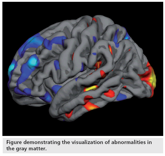
The scan, which was developed at King’s College London (London, UK), could detect small, but crucial, signs of autism in a short 15 min scan. The research, which was led by Christine Ecker, could dramatically improve the time taken to diagnose autism spectrum disorder (ASD) in adults.
The study utilized an MRI scanner and 3D imaging techniques to assess the structure, shape and thickness of the gray matter of the brain. Using a multiparameter classification approach, the team were able to characterize complex and subtle differences in the gray matter of adults suffering from ASD.
A support vector analytical method was used and detected discriminating patterns that can identify an individual with ASD. The results confirm that the neuroanatomy of ASD is multidimensional, and affects multiple neural systems.
Ecker describes the purpose of the research as “working towards establishing neuroanatomy as a biomarker for ASD, which could be used to guide and/or substantiate the conventional behavioral diagnosis in the future”.
Autism is a neurodevelopment condition that has a variety of causes and symptoms. The severity of the condition ranges from mild to severe behavioral symptoms. The condition affects approximately one in 10 0 a du l t s , most of whom are male, and can have severe d e b i l i t a t i n g consequences.
Conventional di a gnos i s of the condition involves assessment by a team of psychologists, who study behavior and response to a series of a s s e s sme n t s . This behavioral diagnosis can take many months and the accuracy of this method in successfully diagnosing ASD is typically approximately 80%.
The research, which was funded by the Medical Research Council, consisted of 20 nonautistic adults, and 20 adults who had previously been diagnosed with the condition. The individuals were originally assessed using the conventional diagnosis method and then subjected to the brain-scan method. The scan method was found to successfully detect ASD in individuals with sensitivity and specificity as high as 90%.
However, Ecker has commented “it is important to mention that we do not propose our approach as a population screening tool, which should be used instead of the conventional diagnosis. Instead, our test could be used to accompany the conventional diagnostic process.”
The study could provide a rapid diagnostic tool based on biological markers for the assessment of autism in adults, and reduce the need for the time consuming and emotional behavior-diagnostic method.
The team are now investigating younger subjects and females that suffer from ASD, and also individuals who suffer from comorbid conditions, such as attention deficit hyperactivity disorder, to see if similar results can be replicated for these groups.
Source: Ecker C, Marquand A, Mourão-Miranda J et al.: Describing the brain in autism in five dimensions – magnetic resonance imagingassisted diagnosis of autism spectrum disorder using a multiparameter classification approach. J. Neurosci. 30(32), 10612–10623 (2010).


