Review Article - Clinical Practice (2022) Volume 19, Issue 1
Laser: Silent revolution in prosthetic dentistry bridging the gap to future review
- Corresponding Author:
- Nagy Abdulsamee
Professor and Chairman of Prosthetic Dentistry Department
Faculty of Dentistry, Deraya University, Egypt
E-mail: nagyabdulsamee@gmail.com
Received: 10 January, 2022, Manuscript No. M-51443 Editor assigned: 11 January, 2022, PreQC No. P-51443 Reviewed: 21 January, 2022, 2022, QC No. Q-51443 Revised: 23 January, 2022, Manuscript No. R-51443 Published: 29 January, 2022, DOI. 10.37532/fmcp.2022.19(1).1833-1848
Abstract
Several technologies have been used to overcome current challenges in dentistry throughout the last few decades. Laser technology is the most recent addition to this group of technologies. Because of its great precision, biocompatibility, and few side effects, it has had a significant impact and has so supplanted several traditional procedures. For the past two decades, lasers have been well-integrated in clinical dentistry, enabling practical choices in the management of both soft and hard tissues, with growing usage in the field of prosthetic dentistry. One of their key advantages is that they can deliver extremely low to extremely high concentrated power at a precise location on any substrate using any method available. New approaches are provided for the development of prosthodontic treatments that demand high energy levels and careful processing, such as metals, ceramics, and resins, as well as time-consuming laboratory processes like cutting restorative materials, welding, and sintering.
Lasers have a wide range of applications, and their use in the field of prosthodontics has seen them replace stainless steel scalpels with optical scalpels to a respectable extent throughout the surgical field and other traditional ways. Most notably, this technique allows dental patients to have intraoral surgery with or without anesthetic, with a higher level of comfort and a faster recovery time. New technology on the horizon will address these flaws, but it will also come with its own set of hazards and restrictions. The purpose of this article is to discuss the application and uses of lasers in prosthodontics, as well as how lasers have revolutionized patient care. A future project could be the development of new intraoral laser devices.
Keywords
laser physics, dental alloys, laser welding, prosthetic dentistry, computer-aided design, laser sintering, esthetic dentistry, implantology, maxillofacial prosthodontics
Laser history
In 1917, scientist Albert Einstein established the theory of stimulated emission, which serves as the scientific foundation for the production of laser light [1]. His theory lay dormant until 1956 when Thomas Maiman created the first laser, the Ruby laser. Mailman exposed an extracted tooth to a prototype Ruby (694 nm) Laser, which resulted in energy transfer. Rubby laser was first employed in 1967 for carrying caries removal and cavity preparation, although it resulted in pulpal damage [2]. The CO2 laser was invented by Patel at Bell Laboratories in 1964 [1], but it was not used in oral surgery until the 1980s for the excision of soft tissue diseases [3].
The Nd:YAG laser was the first laser designed exclusively for dental usage in caries eradication and Kinetic cavity preparation, and it was created in 1987 and approved by the US Food and Drug Administration in 1990 [3]. One year after the Nd:YAG laser, the argon laser was produced. Lasers became widely used in dentistry after the advent of the diode laser. The American Dental Laser, which paved the way for laser dentistry, did not become commercially available until 1989.
Dr. Terry and Bill Meyers invented the first dental laser in 1989, a 3W Nd:YAG for soft tissue application, and since then, a range of laser wavelengths have been introduced and sold [4]. In the mid-1990s, laser dentistry made a breakthrough. In the mid-1990s, dentists were able to choose from a variety of laser types (diode laser, Nd:YAG, Er,Cr:YSGG, Er:YAG, CO2) with appropriate wavelengths (810 nm- 890 nm, 1064 nm, 2780 nm, 2940 nm, 10600 nm respectively).
Introduction
Light is an integral part of our life. LASERS, one of the greatest inventions in science and technology, was developed in the early twentieth century. Light Amplification by Stimulated Emission of Radiation (LASER) was invented by Gordon Gould, a Columbia University doctoral student, in a 1959 study that went on to become a gift to the health sciences [5]. In the medical field, the use of lasers for treatment has become widespread [6]. Lasers are a very appealing technology in numerous sectors of dentistry because of their low intrusive nature and speedy tissue reaction and healing. They serve as a tool to achieve a better result than ever before. The rapid development of lasers and their wavelengths for a variety of soft and hard tissue applications may continue to have a significant impact on prosthetic dentistry’s scope and practice. The goal of this paper is to familiarize every clinician, particularly prosthodontists, with laser fundamentals and different laser systems so that they can incorporate them into their clinical practices [7].
Mechanism of laser production
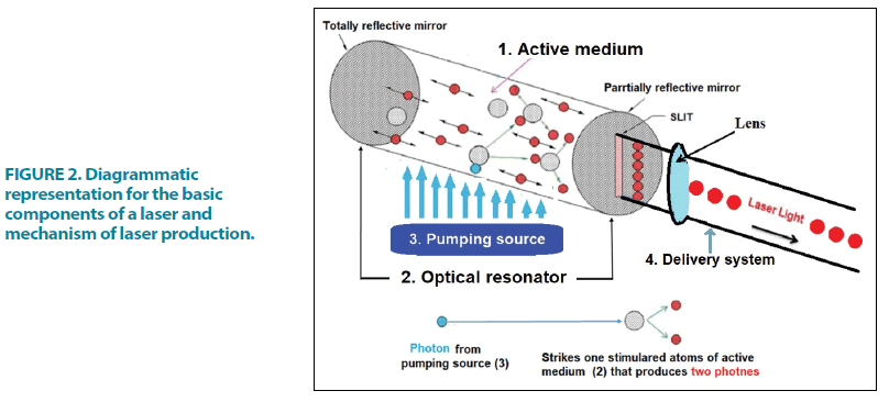
The three separate phenomena that occur during the production of an atomic spectral line outlined by Albert Einstein in 1916 (FIGURE 1 and FIGURE 2) [8] are spontaneous emission, stimulated emission, and absorption.
Amplification of stimulated emission generates laser light [9,10]. Amplification is a step in the laser’s internal process. Understanding how light is produced can be aided by identifying the components of a laser instrument [9-11].
A typical laser is made up of six fundamental components (FIGURE 2).
■ Active medium
A material, either naturally occurring or manmade that when stimulated, emits laser light. This material may be a gas e.g. Argon, CO2), a crystal (YSGG crystal doped with Er and Cr, YAG crystal doped with Er or Nd) or a solidstate semiconductor (AlGaAs, InGaAs diode). The active medium is contained within the laser cavity, which is an internally polished tube with two mirrors, one is completely reflective at one end and the other is partially reflective at the other end and is encircled by the external energizing input, or pumping mechanism. It produces a laser when exited by the pumping mechanism.
■ Pumping mechanism (FIGURE 2)
The pumping unit surrounds the optical cavity, which can be an arc light or a flashlight for excitation, or an electromagnetic coil or diode unit. Its purpose is to provide energy to activate the atoms of the active medium in order to produce laser. The active medium absorbs the energy from the primary source, resulting in the creation of laser light via the stimulated emission process. The phrase “stimulated emission” comes from the quantum theory of physics, which was first proposed in 1900 by German physicist Max Planck and later theorised by Danish physicist Neils Bohr as pertaining to atomic architecture. Due to energy absorption, incident light energy received by a target atom will cause an electron to move to a higher energy shell (FIGURE 1A). In comparison to the stable energy state of the target, this unstable state will result in the emission of photonic energy, with surplus energy being produced as heat. This is referred to as spontaneous emission (FIGURE 1B). Albert Einstein goes on to explain that bombarding an already excited atom with a second photon causes it to emit two coherent photons of the same wavelength, a process he called stimulated emission (FIGURE 1C). Amplification of stimulated emission produces laser light [9,10].
■ Optical resonators
They are usually polished surfaces or two mirrors at each end of the optical cavity that are aligned. The optical resonators are responsible for amplification and collimation of the developing beam.
■ Delivery system
It’s a way to deliver laser energy to a specific location. An articulated arm, a hand-piece, a flexible hollow wave guide, or a quartz fiberoptic are just a few examples.
■ Cooling system
During the creation of laser energy, heat is produced. As a result, a cooling system is employed to keep the compartments cool.
■ Control Panel
The control panel is used to set variable parameters for the laser’s output.
Laser properties
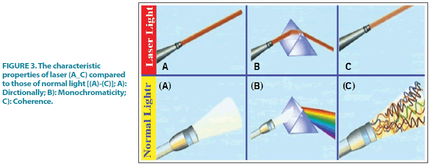
The common characteristics properties of all laser beam types (A-C) in comparison to those of normal light [(A)-(B)] are
■ Directionality
It is a situation in which all waves are parallel to one another and have little divergence or convergence. It allows laser light to travel long distances without loss of intensity (FIGURE 3A). Those of normal light are scattered FIGURE 3A.
■ Monochromaticity
Refers to the fact that it is made up of only one wavelength. This monochromatic feature of laser light has therapeutic significance since it allows it to target certain chromophores like water, hemoglobin, and melanin, allowing for specialized clinical uses (FIGURE 3B). Those of normal light are polychromatic FIGURE 3B.
■ Coherence
All photon waves have the same amplitude and frequency, which is known as coherence. As a result, a special type of focused electromagnetic radiation is produced FIGURE 3C [12]. Those of normal light are non-coherent FIGURE 3C.
Because of laser collimation, monochromaticity, and coherence its Brightness is more intense and brighter compared to the sun. A one mW He-Ne laser, which is a highly directional low divergence laser source, is brighter than the sun, which is emitting radiation isotropically. Brightness is defined as power emitted per unit area per unit solid angle [13].
Classifications of lasers
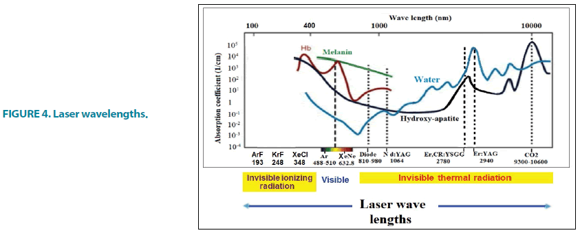
■ According to the wavelength [14]
1. Ionizing lasers (ultraviolet, x-ray, and gamma lasers) (approximately below 400 nm).
2. The visible spectrum range (approximately 400 nm-700 nm).
3. Non-ionizing thermal radiation lasers (the infrared spectrum range (approximately 700 nm to the microwave spectrum).
4. In medical practice, wavelengths ranging from 193 nm to 1060 nm are commonly employed, with wavelengths ranging from ultraviolet to far-infrared (FIGURE 4). Invisible ionizing lasers, on the other hand, are not used in dentistry.
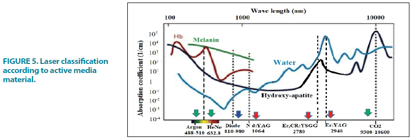
■ According to active medium (FIGURE 5)
It is usual to classify laser devices according to their active material into:
Semiconductor lasers (diode laser): Typically obtained using a p-n junction, pumping with an electrical current. Semiconductor lasers are available in a variety of wavelengths ranging from 810 nm to 980 nm and are versatile enough to be employed for all soft tissue dental procedures.
Gas lasers: These lasers are characterized by gaseous gain media. Pumping is obtained by electrical discharges. The quality of the emitted beam is exceptional. The key characteristic of gas lasers is that they emit efficiently where other types of lasers do not. For example, a CO2 laser with a wavelength of 10.6 μm may output kilowatts of power, making it appropriate for macro-machining, welding, and cutting [15].
Solid-state lasers: Crystals or glasses doped with rare-earth or transition metal ions serve as their active media. Nd:YAG, ErCr:YAG, and Er:YAG are examples of common media. A discharge light is used to pump the water. It is also possible to utilise a diode pump. This type of laser, which emits in the near-IR range, has a wide range of emission power and excellent beam quality.
NB fiber lasers: They operate in a similar way to solid-state lasers in terms of principle. The doped area is found in the fiber’s core. This type of laser has a higher thermal dispersion and the unique ability to operate in transversal single mode [16] due to the fiber shape. This provides for many kilowatts of emission while retaining a greater beam quality [17]. Micromachines, Yb-doped and Yb/Eb-doped fiber lasers are commonly utilized in industrial applications where high precision machining is required. Tm-doped fiber lasers allow for improved contact with soft tissues in medical applications without causing a temperature rise at the operation site. It has the potential to be used for intraoral welding of metallic appliances [18].
■ According to hazards
Certain lasers’ output is dangerous to the human body, and their exposure can harm the eye and skin. Limited access to the room and equipment, the use of personal protective equipment, system monitoring, testing and operations of the laser and its delivery systems, correct applications, and vigilance on the part of each laser team member are all necessary for hazard control [19]. The International Electrotechnical Commission (IEC), a global agency, has developed a new categorization system to identify the amount of laser beam hazard given in TABLE 1 [20].
| Class | Description |
|---|---|
| I | Very low risk, "safe under reasonably foreseeable use" |
| IM | Wavelengths between 302.5 nm and 400.0 nm are safe except when used with optical aids (e.g. binoculars) |
| II | Wavelengths between 400 nm and 700 nm. No known hazard with 0.25 seconds (aversion response) |
| IIM | No known hazard with 0.25 seconds (aversion response) unless collecting optics are used |
| IIIR | Direct intrabeam viewing is potentially hazardous but the risk is lower than Class III B lasers |
| IIIB | Normally hazardous under direct beam viewing conditions, but are normally safe when viewing diffuse reflections |
| IV | Hazardous under both intra beam and diffuse reflection viewing conditions. They may also cause skin injuries and are potential fire hazards |
TABLE 1. Standard IEC Classification.
Laser safety
Any practice that uses lasers must have proper safety protocols in place. The practice should select a laser safety officer whose role is to implement and maintain safety standards. Each device’s maker is required to train providers on the critical precautions that are required for each device. The following are some of the most common safety precautions [19,20]:
■ Eye protection
Protective eyewear for the wavelength in use must be worn by the patient, clinical staff, and any observers.
■ Plume control
Laser operations produce a plume that could contain harmful chemicals and microbes. When utilized properly, standard dental high-speed evacuation is sufficient to control the plume. The clinicians should wear high-quality masks.
■ Sharps
Sharps, such as scored laser tips from quartz fibers, must be disposed of as such.
■ Warning sign
Warning signs must be prominently displayed, and access to the operatory must be restricted.
Laser classification according to emission mode
Any laser emission mode works on the idea that light energy contacts tissue for a specific amount of time, causing a thermal reaction. The dental laser device is capable of emitting light energy in three different modes [21]:
■ Continuous-wave mode
The beam is emitted at only one power level for as long as the operator depresses the footswitch (FIGURE 6a). In this case, the operator must manually turn off the laser so that the tissue can relax thermally.
■ Gated pulse mode
The laser energy is turned on and off on a regular basis, similar to how an eye blinks (FIGURE 6b). The opening and shutting of a mechanical shutter in front of the continuous wave emission’s beam path achieve this mode.
■ Free-running pulsed mode
(“true-pulsed”) This emission is unique in that high peak energies of laser light are emitted for a short time (microseconds), then the laser is turned off for a lengthy time (FIGURE 6c).
Clinical significance of pulse-type
We need a pulsed mode to cut very thin or fragile soft tissue because the amount and rate of tissue removal will be slower, but we need a continuous wave to cut thick dense fibrous tissue because more energy for removal and emission will provide a faster yet safe pace of excision.
Laser tissue interaction
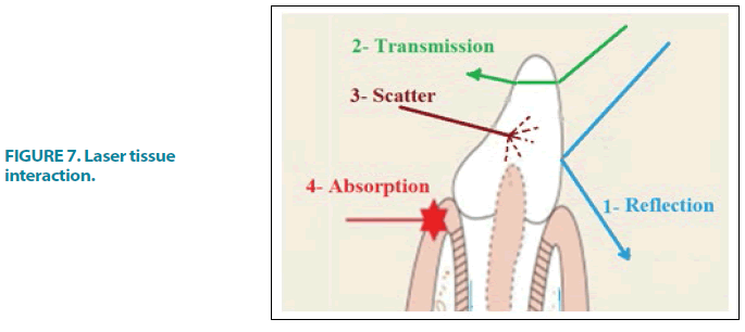
One of four ways light energy interacts with a target medium (e.g. oral tissues) (FIGURE 7) [22]:
■ Reflection
The beam bounces off the tissue surface without penetrating or interfering in any way. Reflection is normally an undesirable phenomenon, however, when Erbium lasers reflect off titanium, it allows for safe gingiva cutting around implant abutments. Because the reflected light may cause injury to the eye, wearing safety eyeglasses before activating the laser is required.
■ Transmission
The laser beam enters the medium and exits distally without interacting with it, causing no damage to the target tissue. H2O is generally clear to Nd:YAG, whereas CO2 is quickly absorbed by cellular fluids. The laser energy can interact with deeper tissues by passing through superficial tissues. For example, in retina surgery, the laser travels through the lens to treat the retina. Tissue transmission is further demonstrated by the deeper penetration obtained with Nd:YAG and diode lasers.
■ Scatter
The laser energy will disperse in many ways once it penetrates the target tissue, resulting in a weakening of the laser energy with no effect on the target tissue. This phenomenon is usually ineffective, although it can aid in the biostimulation qualities of particular wavelengths and the curing of light-activated polymeric materials.
■ Absorption
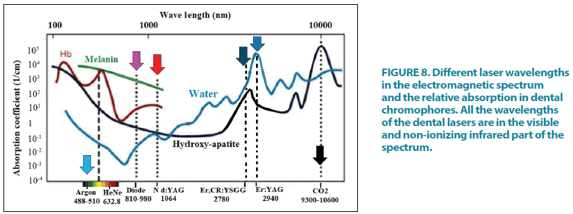
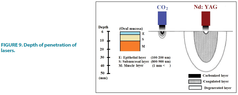
The most significant interaction is absorption, which is the most common favorable outcome. Specific chromophores absorb the energy of each wavelength. This absorbed energy is transformed into thermal and/or mechanical energy, which is then used to complete the task at hand. Pigments like hemoglobin and melanin absorb the majority of near-infrared lasers like diodes and Nd:YAGs. Water and hydroxyapatite absorb the bulk of erbium and CO2 lasers (FIGURE 8). If the incidence wavelength is correct and the delivery parameters are correct, a central zone of tissue ablation is surrounded by a region of irreversible protein denaturation (coagulation, eschar). Along a heat gradient, a reversible, reactive zone of edema will form around it. The depth and degree of this tissue alteration will vary depending on the laser wavelength, with longer wavelengths producing less edema and shorter wavelengths producing more edema (FIGURE 9).
Figure 8: Different laser wavelengths in the electromagnetic spectrum and the relative absorption in dental chromophores. All the wavelengths of the dental lasers are in the visible and non-ionizing infrared part of the spectrum.
Absorption needs chromophores, which are light absorbers with a specific affinity for specific wavelengths of light. The following are dental chromophores (FIGURE 8) [22]:
1. Soft Tissue chromophores like Melanin, Hemoglobin and Water
2. Dental hard tissue chromophores like Water and Hydroxyapatite
3. Photosensitive materials in VLC materials (Camphorquinone)
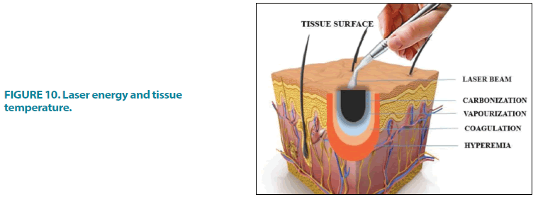
Laser energy and tissue temperature
The initial phase hyperthermia occurs when tissue is exposed to temperatures above normal, but the tissue is not damaged. A temperature of 60ºC causes denaturation and coagulation of the protein. Using the laser parameters within the temperature range, the clinical practitioner can eliminate the granulomatous lesion without vaporizing the surrounding tissue. At 70ºC to 80ºC, the tissue layers adhere to each other. The water within the tissue vaporizes as the temperature reaches 100ºC. At this temperature, the ablation begins, but the dental tooth structures are not ablated. Dehydration is complete at around 200ºC, and the tissue carbonizes (FIGURE 10). Continuous laser treatment causes tissue carbonization, which hinders normal tissue ablation, resulting in tissue necrosis [10,23].
Uses of laser in prosthodontics
■ Fixed prosthodontics
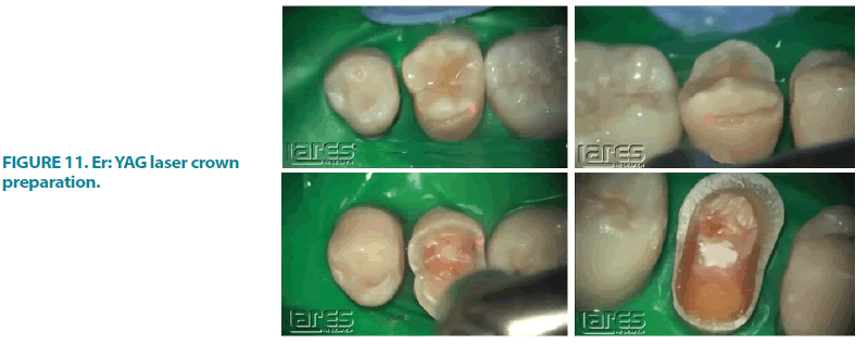
Tooth preparation: When it comes to preparing dental hard tissues, the Er:YAG laser is the recommended treatment. Because the Er:YAG laser numbs the tooth in most cases, anesthesia is rarely required (FIGURE 11). Microfractures in the enamel can be caused by a high-speed handpiece, but there is no chance of microfracture with the laser [24].
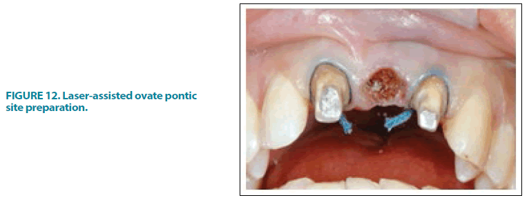
■ Formation of ovate pontic sites (FIGURE 12) [25]
Trough formation: Before the impression operation, a trough is made around the tooth structure. Retraction cord placement, electrocautery, and hemostatic agents are no longer required. During the imprint operation, the laser delivers efficient, predictable, no or limited bleeding, reducing post-operative pain and chair time [26,27]. It also provides coagulation by sealing blood vessels, and it replaces and reduces the usage of hemostatic drugs [28]. The most often used lasers for troughing Nd:YAG, which reduces epithelial attachment interference and postoperative problems.
Laser phototherapy: Low-Level Laser Therapy (LLLT) such as the AsGaAl (gallium aluminum arsenide) 660 nm laser is used in the gingival sulcus and gingival tissue surrounding the crown preparation to promote soft tissue biomodulation, ensuring that no inflammation signals are present in the gingival tissue prior to the final luting procedure [29].
Dentinal decontamination: Due to their superior penetration into dentinal tissues and microbial inhibitory characteristics, High-Level Laser Therapy (HLLT) is employed as a final step before the final cementation of the crown [30-32].
To treat dentinal hypersensitivity: Dentinal hypersensitivity is treated with the Nd:YAG laser [33]. Many lasers can have thermal effects; however, if their characteristics are utilized under controlled conditions, thermal damage to temperature-sensitive pulpal tissues can be reduced.
Veneer removal: The laser energy penetrates through the ceramic glass and is absorbed by the adhesive’s water molecules. At the silaneresin interface, debonding occurs. For ceramic restorations, the method takes around two seconds to two minutes, depending on the thickness of the ceramic restoration [34].
Bleaching: Ar and diode lasers are often used for in-office bleaching treatments. The only laser bleaching system having photothermal, photochemical, and photocatalytic activity is the KTP laser and H2O2 gel combination (Smart Bleach gel [SBI]). The goal of laser teeth whitening is to achieve maximum bleaching power while minimizing side effects [35].
E model preparation: A 3D laser scanner is a useful tool since it is simple to use and does not require cast preparation because e-models are created from scanned impressions. It comprises a laser beam and a camera-like device that uses triangulation techniques [36].
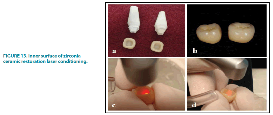
Conditioning of the inner surface of zirconia restoration: The Er:YAG laser was used to irradiate the interior surfaces of the zirconia ceramic restorations (FIGURE 13). The irradiation was carried out for 30 seconds underwater cooling (5 mL/min) at a distance of 12 mm-15 mm from the ceramic surface (enough time for the entire inner surface to be irradiated). For 2 seconds, the surfaces were lightly dried [25].
Crown lengthening: Appropriate crown length is essential for better aesthetics and functional importance and is commonly indicated for teeth with caries at the marginal gingiva level [37], a cuspal fracture that extends below the gingival margin, insufficient clinical crown length, to improve aesthetic value (i.e., in cosmetic use), and when placement of the finish line is difficult. Lasers are used to improve gingival contour and give precise control during surgery. It also provides a dry surgical area for improved vision, bacterial decrease, and faster healing with less time spent in the operating room.
Modification of soft tissues around laminates: The use of an argon laser makes it easier to recontour and remove the residual gingival tissues around laminates [37].
■ Laser use in aesthetic dentistry
Smile design involves the use of lasers such as argon, CO2, diode, erbium, and pulsed Nd:YAG. Soft tissues are pencil-marked to determine the correct gingival shape, symmetry, axial inclination, and zenith before employing the laser. To measure the marginal gingiva, a periodontal probe is positioned apically to the alveolar crest. The biological width is determined and tagged with a laser as a reference place, allowing the physician to concentrate on aesthetics and functioning as a visual completion point [38].
■ The use of laser in removable prosthodontics
The vitality and order of the neighboring tissues in relation are always important factors in the success of the prosthesis placement. A good preprosthetic surgery can increase the success rate and improve the patients’ satisfaction. Lasers are used to treat irregularly resorbed ridges, deformities, and hyperplastic tissue, resulting in changes to both hard and soft tissues for improved function.
Exostoses or tori removal: If they are larger or uneven in shape, or the mucosal covering gets ulcerated, it might lead to prosthetic issues. These may interfere with the mandibular prosthesis’ lingual extension or flanges, or the maxillary prosthesis’ palatal coverage. The treatment of choice for removing bony protuberances is Er:YAG. The main benefit of using an Er:YAG laser is that it reduces overheating, decontamination, biostimulation, and the formation of a smear layer, which obstructs the healing process [39].
Treatment of unsuitable alveolar ridges: Illfitting dentures increase the pressure on the natural residual tissue structures, which are not completely handled by soft linings, because of the lower stress carrying surface. Soft tissue lasers are now commonly utilized in surgery to reveal hard tissue-like bone using a variety of soft tissue wavelengths. For hard tissue treatment, Nd:YAG lasers, CO2 laser diodes, and Erbium lasers are commonly used [40].
Vesitbuloplasty: Vestibuloplasty is used when prosthesis stability is compromised by mandibular or maxillary crest atrophy or a short vestibule. CO2 technology is a straightforward and safe solution. Sutures and grafts are unnecessary. Removable dentures must be relined with soft acrylic right away and only temporarily [41].
Fibroma: Fibrous tissue can form as a result of a sharp denture flange or over-compression of the posterior palatal seal area, causing persistent tissue injury. Any soft tissue laser can be used to treat this, allowing the tissue to re-epithelialize. To promote a faster and higher quality of healing, carbonization was simply eliminated with a cotton swab dipped in saline solution [42].
Epulis fissuratum: Epulis is an overgrowth of the mucosa produced by prolonged tissue irritation in complete denture-wearing patients. Soft tissue lasers, surgical or electrical scalpels are the most often utilized treatments for removing the lesion [43].
Frenectomy: The presence of a high labial frenulum (median or laterals) might cause the oral prosthetic structure to become unstable, and it should be removed to improve denture retention. A thin layer of soft tissues covering the maxillary bone up to the end of the vestibule should be maintained. The frenulum insertion at the level of the lip should be removed as well. Only the lip’s side of the frenulum was sutured [40].
Treatment of undercut alveolar ridge: A mild undercut on both sides may help the denture retain its shape, but a significant undercut on both sides may compromise the denture’s retention and stability. Undercut alveolar ridges are caused by an enlarged tooth socket that is poorly compressed to cover it. Natural undercuts are most typically visible in the lower anterior region or in the case of predominately premaxillary regions. As a result, undercut areas should be surgically addressed before prosthesis management [28]. The Er family of lasers are used for osseous surgery and hard tissue recontouring, while CO2, diode, and Nd:YAG lasers are used for soft tissue treatment [28].
Treatment of enlarged tuberosity: Unopposed maxillary molar teeth cause hyperplastic tuberosity, which may rest against the palate, as well as hyperplastic soft tissues [44]. In such circumstances, prosthetic treatment may result in inappropriate denture placement, poor retention, and stability, as well as pain due to increased load toward a specific location. As a result, pre-prosthetic surgery should be performed to control the condition, and advances in the use of soft tissue and hard tissue lasers may improve prosthetic success rates by allowing for faster wound healing.
Denture fabrication: Selective Laser Sintering (SLS) is used to create dentures. Laser fusion uses a CO2 laser with a high power beam to melt polymer or metal powders at a high temperature. As a result, it creates the strongest and most long-lasting design from a digitized model stored in the Standard Triangulation Language (STL) format [45].
Denture stomatitis: Denture stomatitis is a persistent candidal infection that affects 60 to 65 percent of those who wear dentures. The laser beam not only aids in the ablation of superficial candida-infested epithelial surfaces but also helps to prevent irritation of normal mucosa nearby. Antibiotics or nonsteroidal antiinflammatory medicines will not be needed after the procedure because the laser is virucidal and bactericidal. This may help to avoid bacterial reinfection or secondary infection. Due to the neuron sealing action, lasers aid inappropriate pain alleviation [46].
Investigating the accuracy of impression and complete denture occlusion: The newly created laser scanner is a 3D digitizer that tracks the specimen’s coordinates (x,y,z) and records the information with a resolution of 130 mm at 100 mm. Complex 3D texture-mapped models are captured by the 3D laser and transferred to a 3D (Scan Surf ) software tool, where they are constructed and triangulated into a 3D meshwork image of the object [47]. A 3D model is always preferable to a 2D model. Without contacting the objects, the laser scanner features a 3D Digitizer. As a result, precise data is recorded and saved. The 3D digitizer is linked to the software and the design picture, allowing for the fabrication of an accurate prosthesis. Landmarks on 3D models serve as a guide for fabrication, allowing for speedier tracking and the creation of a standardized prosthesis [48].
Prototyping and CAD/CAM technology: Rapid prototyping can create physical models of CAD data automatically. As a result, it functions as a 3D printer that can aid in the creation of precise prostheses. Using CAD/CAM, this technology created a complete titanium denture base in a short amount of time. Laser scanners, common software, and a prepared denture base plate with a number code. The titanium denture base plate is built up layer by layer [48].
■ Maxillofacial prosthodontics
3D Printing: 3D printing for both bony and soft tissue reconstruction, has become the essential clinical tool that derives a model from a Computer-Aided Design (CAD) built in a layerby- layer fashion. Various 3D printing techniques have been explored in the clinical setting such as stereo-lithography, multi-jet modeling, SLS, binder jetting, and fused deposition modeling [49,50].
■ Laser use in implantology
Second stage uncovering: Overlying soft tissue is removed using CO2 lasers and almost all other types of lasers. Due to low blood contamination and tissue shrinkage, an immediate impression can be taken after the second stage surgery.
Repair of ailing implant: After 1 minute of toluidine blue O implantation, LLLT such as diode soft laser (690 nm) is employed on the contaminated surface for 60 seconds. It lowers the number of microorganisms by 92 percent.
Implant site preparation: The removal of the overlaying structure at the implant site promotes faster healing, better integration, less patient discomfort, and more bone-to-implant contact [51].
Removal of diseased tissue around the implant: In cases of inflammation, lasers are used to remove granulation tissue from the Osseointegrated surface of the implant. Laser energy can be used to sterilize the implant surface. For sterilizing implant surfaces, diode, CO2, and Er:YAG lasers are routinely employed [6], eliminating bacterial contamination and enhancing the success rate [52]. The Er:YAG Laser can be used to remove subgingival calculus from the surface of a titanium implant fixture without causing thermal damage [6].
Lasers for welding prosthetic appliances: In clinical dentistry, cutting the metal framework after casting is common due to a poor fit in implant or dental abutments. The most common reason is wax pattern distortion, as well as deformation or strains during casting [53]. Welding the split pieces of the metal framework has been employed to overcome laboratory distortion problems that result in metal framework mismatch in the marginal area. Welding the metal pieces decreases framework deformation and enhances fit to the abutments as well as stress distribution uniformity [53].
Connecting the metal framework’s sections has been done in a variety of ways. Laser-assisted welding is the favored method among them because it allows the alloy to melt and integrate into the original framework by forming a new joint [53,54]. When a light beam strikes the metal’s surface, the metal absorbs it and converts it to heat, which then penetrates the metal due to conductivity. Because of the high heat concentration, the metal melts and forms a hollow cavity that will be filled with molten metal [55].
This technological achievement has been made even more efficient by the industrial enhancement of compact powerful units of pulsed Nd:YAG lasers. Due to the concentration of energy in a relatively tiny portion of the framework, laser-assisted welding is precise and well defined. In addition, the operator has complete control over each stage of the welding process at all times. Any distortion that occurs during the welding process can be identified and repaired instantly by placing fresh spot welds on the opposite side of the original distortion point [56]. Laser welding appears to give precision, reduced distortion, and a small heat-affected zone in the dental lab [57].
In a case study involving intraoral welding with an Nd-YAG laser, a metallic (Co-Cr-Mo) bar for a maxillary overdenture was welded intraorally in 47 seconds with no pain or discomfort reported by the patient [58]. The Nd-YAG laser is a good choice for welding orthodontic wires in orthodontics [59].
Laser welding component of removable partial dentures: For welding fractured metallic framework, a Nd:YAG laser is employed (FIGURE 14). In comparison to soldering, laser welding can produce 20 percent to 50 percent greater tensile strengths [60].
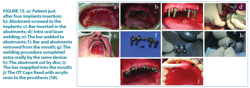
Intraoral Laser Welding (ILW) in implant prosthetic dentistry [58]: A 67-year-old man visited our office for a physical examination. The patient seemed edentulous on the upper arch, where he had a total removable prosthesis, during the clinical examination. His issue was that the denture was not stable, and he found it difficult to speak and eat. The choice was made to put four implants in (FIGURE 15a). Four abutments were screwed to the implants after the surgical operation (FIGURE 15a and FIGURE 15b).
Figure 15: a): Patient just after four implants insertion; b): Abutment screwed to the implants; c): Bar inserted in the abutments; d): Intra oral laser welding; e): The bar welded to abutments; f): Bar and abutments removed from the mouth; g): The welding procedure completed extra orally by the same device; h): The abutment cut by disc; i): The bar reapplied into the mouth; j): The OT Caps fixed with acrylic resin to the prosthesis [58].
Then, a bar previously constructed by the dental technician previewing the position of the implants by the maxillary arch impression was fitted in the four abutments (FIGURE 15c). The bar was welded intraorally (FIGURE 15d) using a dental Nd:YAG laser to secure the position. The entire intraoral welding treatment lasted 47 seconds, and the patient reported no pain or discomfort during it (FIGURE 15e).
The bar was withdrawn from the mouth with the abutments (FIGURE 15f), the welding technique was completed extra orally with the same device (FIGURE 15g), the abutments were cut and polished, and it was then reintroduced into the mouth (FIGURE 15h and FIGURE 15i). Four silicon OT Cap (Rhein 83, Italy) were then used to secure the prosthesis to the bar (FIGURE 15j). After two days, seven days, and fifteen days, the patient was evaluated again, then monthly for six months, and no concerns were found.
■ 3D printing in digital prosthetic dentistry
Metal frames for metal-ceramic restorations are made using laser sintering. Its goal is to eliminate the negative effects of casting while also lowering the financial cost and labor time. The porosity of the material, the fit in the marginal area, and the mechanical qualities of the material, in general, are critical to the reconstruction’s effectiveness. Selective laser sintering is a layered manufacturing technique that allows complicated three-dimensional items to be created by hardening consecutive layers of material powder on top of one another. It is accomplished by employing the heat energy generated by a concentrated laser beam to treat the targeted locations. Each layer is scanned depending on the individual cross-section, as determined by the CAD model, using a beam deflection system (galvanic mirrors). A powder deposition technique is used to deposit consecutive layers of material (ranging in thickness from 20 m to 150 m) [61,62].
■ Laser sintering of crowns and bridges made from non-precious alloys
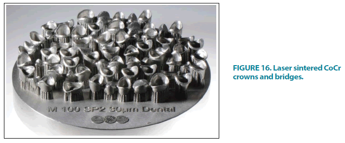
For the fabrication of Co-Cr crowns and bridges, laser sintering has now become the industry standard [63]. It is now possible to create fully stress-free and properly fitted nonprecious metal frames even for bigger bridge spans by optimizing post-processing after the actual building process. Because of the high number of units that can be placed on a single platform, the manufacturing time per unit has been lowered to just a few minutes (FIGURE 16). When it comes to fixed restorations composed of non-precious alloys, the process is incredibly cost-effective and well-established.
■ Laser sintered of clasp retained cast metal frameworks
One-piece prostheses can now be planned digitally and manufactured subtractively using CNC milling units or additively using 3D printing thanks to the introduction of digital techniques for denture production, such as Computer-Aided Design/Computer-Aided Manufacturing (CAD/CAM) and additive manufacturing techniques [64].
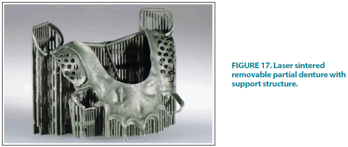
The difference between indirect and direct fabrication processes may be seen here. The frameworks are printed in wax or polymers and then cast using the lost-wax technique in the indirect approach. The CAD data set is immediately turned into a Co-Cr alloy object by laser sintering in the direct approach (FIGURE 17) [65]. This approach is still in its early stages of development. In recent articles, laser sintering has been reported to provide advantages in digital manufacturing in terms of standardization, shorter production times, and easy digital data transfer [66]. Its economic viability, however, is currently being evaluated [67]. Before this procedure can be confidently suggested, more research is required. The retaining elements (clasps) require special attention since they are constantly exposed to significant mechanical loads while performing their retaining and supporting functions.
The advantages of laser sintering include the rapid building of complicated metal frameworks and the use of an automated system, as well as the reduction in working time due to the elimination of numerous phases such as wax model making, coating, dewaxing, and casting. Because the required amount of the alloy is accurately selected and regulated, it saves on the use of the alloy by decreasing metal waste. The success of a dental restoration depends on how well the restoration fits in the marginal area. An in vitro study compared marginal fit and axial wall adaptability of cobalt-chromium copings fabricated using either a conventional lost-wax or a metal laser sintering technique, finding that the laser sintering technique had significantly higher marginal fit and axial wall adaptability than the lost-wax technique, though marginal fit values for both techniques were within clinically acceptable limits [62]. Laser sintering is a technology that requires expensive equipment, which is one of its drawbacks.
Future perspectives
Due to expensive installation costs and inadequate control over the thermal effect of lasers on soft/hard tissues, laser use was primarily limited to professionals and researchers. Dentists are more motivated to use lasers in their clinical practice as a result of recent advancements in laser technology and lower costs. Dentists may perform laser applications on their patients with much more safety than before, owing to smaller laser pulses and lower energy. Lasers are a very appealing tool for the dental industry because of their minimally invasive nature and increased tissue response with better recovery. The difficulty of setting the parameters in relation to the material utilized, its thickness, the gap dimension, and the filler alloy are the most challenging and problematic aspects of using laser technology for dental welding. Laserassisted welding has almost completely replaced traditional soldering processes among dental workers. In the near future, laser sintering-based metal framework construction may be able to replace waxing and casting. Furthermore, Nd:YAG laser welding equipment has traditionally been big and expensive to install and maintain. As a result, the development of simple, inexpensive machines that are simple to operate and require little training would be a fantastic opportunity for dentists to weld metals directly in their offices [68].
The wavelength, power, intensity, fluency, and pulse duration of the many laser systems available for intraoral and laboratory application vary. Although a variety of devices are available for dental applications, some industrial lasers appear to be more effective in the processing of very hard materials, such as zirconia. The ability of ultrafast lasers to ablate and cut the Yttria Stabilised Zirconia (YTZP) surface with excellent precision, creating a wide variety of spots or lines, justifies additional exploration. Such laser equipment is expensive, which is a major drawback that limits its use in dental laboratories. Furthermore, the cost-effective manufacture of intraoral ultrafast laser handpieces (of minimal size) appears to be a future goal [69].
Conclusion
Shorter and painless procedures, ranging from soft tissues to hard tissues, are possible using laser, with low to no discomfort and hence less harm to the surrounding tissue. For its major benefits in dentistry, clinicians should have a thorough understanding of basic laser physics, characteristics, and tissue interactions. For the greatest results in prosthetic dentistry, proper training is required for operating lasers precisely with the use of appropriate wavelengths.
One of the most current is laser welding, which is capable of producing high-quality weld connections with exceptional consistency. More advancements could include combining diagnostic and therapeutic laser welding technologies in one device for finer laser welding operations, robotic aided dental laser welding devices could be useful. It will not be remarkable if, by the middle of this century, laser welding technology has superseded most conventional options.
It is feasible to safely and effectively repair metals intraorally with a dental Nd:YAG laser without putting the patient in danger or causing discomfort, therefore avoiding the costly and time-consuming process of impression taking with its inherent errors.
Ethical Considerations
Not applicable.
Conflict of Interests
None.
References
- Aoki A, Sasaki KM, Watanabe H, et al. Lasers in nonsurgical periodontal therapy. Periodontol 2000. 36, 59-97 (2004).
- Coluzzi DJ. Fundamentals of dental lasers‑ science and instruments. Dent Clin North Am. 48, 751‑770 (2004).
- Frame JW. Carbon dioxide laser surgery for benign oral lesions. Br Dent J. 158, 125-128 (1985).
- Thoma D. Lasers in Prosthetics. Indian J Dent Adv. 6, 1600-1604 (2014).
- Gross AJ, Herrmann TR. History of lasers. World J Urol. 25, 217‑220 (2007).
- Gosawi S, Kumar S, Lakhyani R, et al. Lasers in prosthodontics-A review. J Evol Med Dent Sci. 1, 624‑633 (2012).
- Gounder R, Gounder S. Laser science and its applications in prosthetic rehabilitation. J Lasers Med Sci. 7, 209-213 (2016).
- Welch AJ, Torres JH, Cheong WF. Laser Physics and Laser-Tissue Interaction.Tex Heart Inst J. 16, 141-149 (1989).
- Coluzzi DJ. An overview of laser wavelengths used in dentistry. Dent Clin N Am. 44, 753-765 (2000).
- Mercer C. Lasers in dentistry: a review. Part I. Dent Update. 23, 74-80 (1996).
- Katzir A. Lasers and optical fibers in medicine. 1st ed. Academic Press, USA. 317 (1993).
[Google Scholar] [Crossref]
- Singh SC, Zeng H, Guo C, et al. Lasers: fundamentals, types, and operations. (2012).
[Google Scholar] [Crossref]
- Singh SC, Zeng H, Guo C, et al. Nanomaterials: processing and characterization with lasers. 1st ed. (2012).
[Google Scholar] [Crossref]
- Weber MJ. Handbook of Laser Wavelengths. Ed. Marvin J. Weber Boca Raton: CRC Press LLC. (1999).
[Google Scholar] [Crossref]
- Zhang Y, Killeen T. Gas Lasers: CO2 Lasers-progressing from a varied past to an application-specific future. Laser Focus World. 4, (2016).
- Molardi C, Poli F, Rosa L, et al. Mode discrimination criterion for effective differential amplification in Yb-doped fiber design for high power operation. Optics Express. 25, 29013-29025 (2017).
- Jauregui C, Limpert J, Tünnermann A. High-power fibre lasers. Nature Photon. 7, 861-867 (2013).
- Molardi C, Sun B, Yu X, et al. Polarization-maintaining large mode area fiber design for 2 μm operation. IEEE Photonics Tech Letters. 28, 2483-2486 (2016).
- Smalley PJ. Laser safety: risks, hazards, and control measures. Laser Ther. 20, 95-106 (2011).
- Bargman H. Laser Classification Systems. J Clin Aesthet Dermatol. 3, 19-20 (2010).
- Bhat AM. Lasers in prosthodontics-An overview part 1: fundamentals of dental lasers. J Indian Prosthodont Soc. 10, 13-26 (2010).
- Gupta S, Kumar S. Lasers in dentistry-an overview. Trends Biomater Artif Organs. 25, 119-123 (2011).
- Russel AD. Lethal effects of heat on bacterial physiology and structure. Sci Prog. 86, 115-137 (2003).
- Fred M. Erbium laser.J Can Dent Assoc. 70, 334-335(2004).
[Google Scholar] [Crossref]
- Dyer B. Minimally invasive osseous crown-lengthening procedure using an erbium laser: clinical case and procedure report. J Cos Dent. 23, 72-78 (2008).
[Google Scholar] [Crossref]
- Parker S. The use of lasers in fixed prosthodontics. Dent Clin N Am. 48, 971-998 (2004).
- Lukram A, Sachdeva N, Sahu K, et al. Application of laser in prosthetic dentistry-A review. Int J Dent Med Res. 1, 99‑102 (2014).
- Durrani S. Lasers and it’s application in prosthetic dentistry. Int J Dent Med Res. 1, 183‑188 (2015).
- Cardoso Bezerra SJ, Fioranelli Vieira G, Eduardo Cde P, et al. Laser phototherapy (660 nm) can be beneficial for reducing gingival inflammation in prosthodontics.Case Rep Dent. 2015, 132656 (2015).
- Pejcic A, Kojovic D, Kesic L, et al. The effects of low level laser irradiation on gingival inflammation. Photomed Laser Surg. 28, 69-74 (2010).
- Ramalho KM, de Freitas PM, Correa-Aranha AC, et al. Lasers in esthetic dentistry: soft tissue photobiomodulation, hard tissue decontamination and ceramic conditioning. Case Rep Dent. 2014, 927429 (2014).
- Klinke T, Klimm W, Gutknecht N. Antibacterial effects of Nd:YAG laser irradiation within root canal dentin. J Clin Laser Med Surg. 15, 29-31(1997).
- Malhotra R, Thurkral H. Laser application in prosthodontics-A review. Int J Enhanc Res Med Dent Caries. 3, 20-25 (2016).
[Google Scholar] [Crossref]
- Spitz SD. Lasers in prosthodontics: clinical realities of a dental laser in a prosthodontic practice. Alpha Omegan. 101, 188-194 (2008).
- Roeland JG. The use of KTP laser, an added value for tooth bleaching. J Oral Laser App. 9, 219-226 (2009).
- Guth JF, Keul C, Stimmelmayr M, et al. Accuracy of digital models obtained by direct and indirect data capturing. Clin Oral Investig. 17, 1201-1208 (2013).
- Kalra T, Magrath M, Kalra G. Lasers in prosthodontics-Part I implantology. Int J Laser Dent. 4, 49‑53 (2014).
- Adams TC, Pang PK. Lasers in aesthetic dentistry. Dent Clin N Am 48:833-860 (2004).
- Rocca JP. Er:YAG laser: a new technical approach to remove torus palatinus and torus mandibularis. Case Rep Dent. 2012, 487802 (2012).
- Kesler G. Clinical applications of lasers during removable prosthetic reconstruction. Dent Clin N Am. 48, 963-969 (2004).
- Kumar R, Jain G, Dhodapkar SV, et al. The comparative evaluation of patient’s satisfaction and comfort level by diode laser and scalpel in the management of mucogingival anomalies. J Clin Diagn Res. 9, ZC56-ZC58 (2015).
- Namour S. Atlas of current oral laser surgery. Boca Raton, FL: Universal Publishers. (2011).
- de Arruda Paes-Junior TJ, Cavalcanti SC, Nascimento DF, et al. CO2 laser surgery and prosthetic management for the treatment of epulis fissuratum. ISRN Dent. 2011, 282361 (2011).
- Das M, Paul R, Ali M, et al. Laser in prosthodontics. Univ J Dent Sci. 2, 9-12 (2017).
[Google Scholar] [Crossref]
- Wu J, Gao B, Tan H, et al. A feasibility study on laser rapid forming of a complete titanium denture base plate. Lasers Med Sci. 25, 309-315 (2010).
- Keerthana S, Sivakumar P, Sudhakar R, et al. Denture stomatitis treatment with diode laser. Intern J Prosthodontics Restor Dent. 1, 55-57 (2011).
- Sandesh G, Sanjay K, Rohit L, et al. Lasers in Prosthodontics.J Evol Med Dent Sci. 1, 624-633 (2012).
- Devi N, Kumar PA, Rakshna M, et al. Application of lasers in prosthodontics: A review. J Indian Acad Dent Spec Res. 5, 42-45 (2018).
- Tukuru N, Gowda KP, Ahmed SM, et al. Rapid prototype technique in medical field. Res J Pharm Tech. 1, 341-344 (2008).
- Subburaj K, Nair C, Rajesh S, et al. Rapid development of auricular prosthesis using CAD and rapid prototyping technologies. Int J Oral Maxillofac Surg. 36, 938-943 (2007).
[Google Scholar] [Crossref]
- Baidey SK, Singhal MK, Kumar A, et al. Uses of lasers in prosthodontics. J Dent Sci Oral Rahabil. 6, 125‑127 (2015).
[Google Scholar] [Crossref]
- Gupta R, Luthra RP, Gupta S. Lasers in dental implantology-A review. Int Dent Med J Adv Res. 2, 1‑4 (2016).
- Perveen A, Molardi C, Fornaini C. Applications of laser welding in dentistry: A state-of-the-art review. Micromachines. 9, 209 (2018).
- Bertrand C, Le Petitcorps Y, Albingre L, et al. The laser welding technique applied to the non-precious dental alloys: Procedure and results. Br Dent J. 190, 255-257 (2001).
- Tambasco J, Anthony T, Sandven O. Laser welding in the dental laboratory: An alternative to soldering. J Dent Technol. 13, 23-31 (1996).
- Bertrand C, Poulon-Quintin A. Proposals for optimization of laser welding in prosthetic dentistry. J Prosthodont. 19, 69-76 (2010).
- Varghese S, Bhat V, Joseph S. Laser welding of dental alloys: A systematic overview. Libyan Dent J. 4, 17217481 (2014).
- Fornaini C, Merigo E, Cernavin I, et al. Intraoral Laser Welding (ILW) in implant prosthetic dentistry: Case report. Case Rep Dent. 2012, 839141 (2012).
- Jain S, Vibhute PK, Patil C, et al. Laser welding in orthodontics: A review study. J Dent Health Oral Res. 1, 1-14 (2020).
- Carek A, Zivko-Babic J, Schauperl Z, et al. Macroscopic analysis of Co-Cr base alloys joints. Acta Stomatologica Croatica. 41, 216-224 (2007).
- Kruth JP, Mercelis P, Vaerenbergh JV, et al. Binding mechanisms in selective laser sintering and selective laser melting. Rapid Prototyp J. 11, 26-36 (2005).
- Gaikwad BS, Nazirkar G, Dable R, et al. Comparative evaluation of marginal fit and axial wall adaptability of copings fabricated by metal laser sintering and lost-wax technique: An in vitro study. J Indian Prosthodont Soc. 18, 47-52 (2018).
- Revilla-Leon M, Meyer MJ, Ozcan M. Metal additive manufacturing technologies. Int J Comput Dent. 22, 55-67 (2019).
- Schweiger J, Kieschnick A. CAD/CAM in digital dentistry; Teamwork Media: Fuchstal, Germany. (2017).
[Google Scholar] [Crossref]
- Alifui-Segbaya F, Williams RJ, George R. Additive manufacturing: A novel method for fabricating cobalt-chromium removable partial denture frameworks. Eur J Prosthodont Restor Dent. 25, 73-78 (2017).
- Tregermann I, Renne W, Kelly A, et al. Evaluation of removable partial denture frameworks fabricated using 3 differnet techniques. J Prosthet Dent. 122, 390-395 (2019).
- Van Zeghbroeck L, Boons E. Evaluation of technicians working time in the fabrication of removable partial dentures: Cad/Cam versus tradition. In proceedings of the 14th Biennial Meeting of the International College of Prosthodontics, Waikoloa Village, HI, USA. 63 (2011).
- Fornaini C, Sozzil M, Merigo E, et al. Supercontinuum source in the investigation of laser-tissue interactions: Ex vivo study. J Biomed. 2, 12-19 (2017).
- Tzanakakis E-GC, Skoulas E, Pepelassi E, et al. The use of lasers in dental materials: A review. Materials. 14, 3370 (2021).

