Clinical images - Imaging in Medicine (2017) Volume 9, Issue 6
Cardiac amyloidosis revealed by bone scintigraphy
Yassir Benameur1*, Maryam Nadif2, Imad Ghfir1, Latifa Oukerraj2, Hasnae Guerrouj1, Mohamed Cherti2, Nouzha Ben Rais Aouad11Department of Nuclear Medicine, Ibn Sina Hospital, Mohammed V University of Rabat, Rabat, Morocco
2Department of Cardiology B, Ibn Sina Hospital, Mohammed V University of Rabat, Rabat, Morocco
- Corresponding Author:
- Yassir Benameur
Department of Nuclear Medicine
Ibn Sina Hospital, Mohammed V University of Rabat, Rabat, Morocco
E-mail: benameur.yassir@gmail.com
Abstract
amyloidosis ▪ bone scintigraphy
A 61 year old male with cardiovascular risk factors including age, diabetes, dyslipidemia and chronic smoking weaned 4 years ago, who also had a history of implantation of a double chamber pacemaker in 2014, hospitalized several times for acute decompensated heart failure with left ventricular (LV) dysfunction.
The patient was admitted to the hospital for a sudden loss of consciousness. Physical examination revealed a blood pressure at 135/80 mmHg, a normal pulse rate and objectified muffled heart sounds.
The chest X-ray film did not find enlargement of the heart or pleural effusion and a routine electrocardiogram found a left bundle branch block.
Transthoracic echocardiogram showed a left ventricular hypertrophy with severe global hypokinesia and LV dysfunction (ejection fraction: 35%). Biological evaluation was negative. The patient underwent a bone scintigraphy, since in his case cardiac magnetic resonance imaging was contraindicated (pacemaker device).
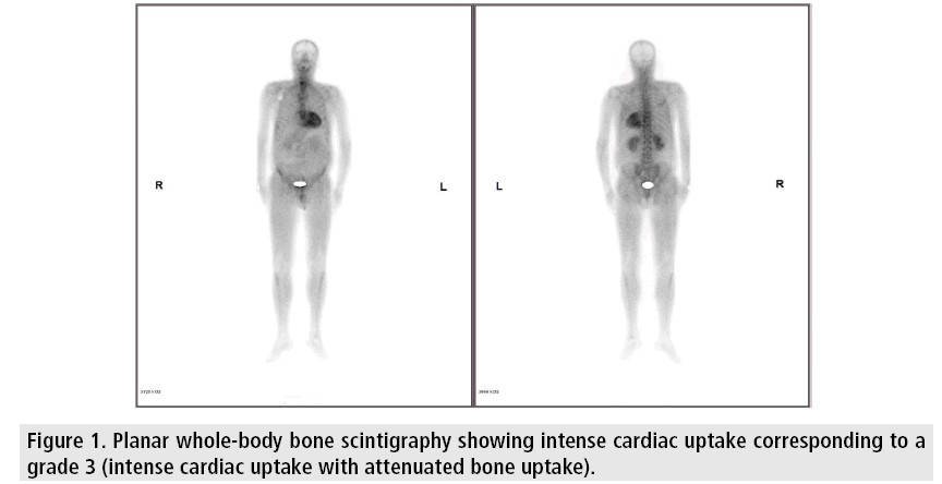
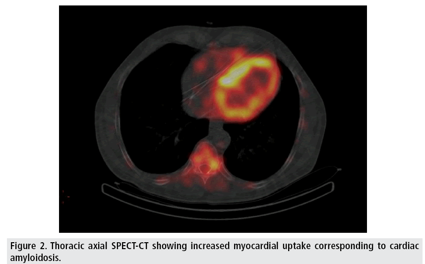
Planar whole-body bone scan was performed in the anterior and posterior projections 3 hrs after injection of 740 MBq (20mCi) of 99mTc- HMDP (99m Technetium hydroxymethylene diphosphonate). The scanning speed was at 10-15 cm/min and the image format was 1024 × 256. Single photon emission computed tomography (SPECT-CT) was additionally performed. The bone scintigraphy showed an intense cardiac uptake corresponding to a grade 3 (intense cardiac uptake with attenuated bone uptake) and attenuated tracer uptake throughout the skeleton (FIGURES 1 and 2).
Figure 1: Planar whole-body bone scintigraphy showing intense cardiac uptake corresponding to a grade 3 (intense cardiac uptake with attenuated bone uptake).
We retained in front of the negative immunological assessment and the bone scintigraphy findings, the diagnosis of cardiac amyloidosis. Our patient has been taken out of biopsy, with its attendant risks and cost and which is not yet feasible in Morocco. Several scintigraphic tracers allow the visualization of amyloid deposits, but the most used are the bone tracers (99mTc-DPD, 99mTc-HMPD, 99m Tc- MPD, 99m Tc-PYP) [1].
They bind to cardiac amyloid deposits with a very good sensitivity for transthyretin amyloidosis (hereditary or senile), while AL amyloidosis absorbs these tracers only slightly. An intense cardiac uptake on bone scintigraphy is therefore in favor of a transthyretin amyloidosis, while a negative scintigraphy cannot eliminate AL amyloidosis [2]. Gillmore et al. have concluded that cardiac transthyretin amyloidosis can be reliably diagnosed in the absence of histology provided that all of the following criteria are met: Heart failure with an echocardiogram or cardiac magnetic resonance that is consistent with or suggestive of amyloidosis, grade 2 or 3 cardiac uptake on a radionuclide scan with 99mTc-DPD, 99mTc-PYP, or 99mTc-HMDP and absence of a detectable monoclonal gammapathy [3], which was the case of our patient.
In conclusion, this report underlines the importance of bone scintigraphy in the detection of cardiac transthyretin amyloidosis, particularly in our context where endomyocardial biopsy is not yet realized.
References
- Lairez O, Pascal P, Victor G, et al. Bone scintigraphy for cardiac amyloidosis imaging: Past, present and future. Med Nucl. 41, 108-114, (2017).
- Bodez D, Galat A, Guellich A, et al. Les amyloses cardiaques: les reconnaître et les prendre en charge. Presse Med. 45, 845-855, (2016).
- Gillmore JD, Maurer MS, Falk RH, et al. Non-biopsy diagnosis of cardiac transthyretin amyloidosis. Circulation. 133, 2404-2412, (2016).


