Case Report - International Journal of Clinical Rheumatology (2020) Volume 15, Issue 3
B-mode ultrasound in the uveitis in the psoriatic arthritis without skin lesion
- Corresponding Author:
- José Alexandre Mendonça
1Serviço de Reumatologia
Pontifícia Universidade Católica de Campinas
São Paulo, Brazil
E-mail: mendoncaja.us@gmail.com
E-mail: Arina.Bingeliene@uhn.ca
Abstract
Introduction: The involvement of inflammatory ocular and joint manifestations can be better evaluated by ultrasound, but the application of this image method in the eyes is a universe that is still very little explored in inflammatory joint diseases.
Objective: The aim of this case report was to detect inflammatory ocular and joint changes by ultrasound with a high frequency linear probe in patients without prior established diagnosis. Case report: This case describes a caucasoid patient of the female sex, 32 years of age, without diagnosis. The patient referred red eyes when using a computer, with weak nails and left ankle arthritis, for 6 months. An ultrasound with 12 MHz high frequency linear probe was used to evaluate the eyes and a frequency of 15 to 18 MHZ for joint investigates.
Discussion: The ultrasound showed in eyes the following findings: floating hypoecogenic images in vitreous humor more intensely to the left, characterizing vitritis, in all of the distal interphalangicas evaluated, with loss of the plate nail pattern trilaminar, effusion tibio - talar, tenosynovitis peroneus longus tendon and entesopathy Achilles tendon, without the presence of power Doppler.
Conclusion: B-mode ultrasound can an important adjunct to the clinical evaluation in patients with arthritis e uveitis. Future studies will better signal the intensity of these findings of vitreous humor alterations, and also define this relationship with the subclinical and inflammatory, ocular and joint activity condition.
Keywords
ultrasound • uveitis • vitritis • arthritis
Introduction
Intermediate uveitis is an often insi dious type of uveitis that occurs mostly in young patients, inflammatory cells in the anterior vitreous, inferior pars plana white exudate, and frequent late cystoid macular edema are the clinical hallmarks for the diagnosis of intermediate uveitis. Not uncommonly, ocular structures, which cannot be seen with routine examination methods. Ultrasound can provide excellent pictures of anterior uvea, vitreous base, and peripheral retina in normal eyes and also in eyes with intermediate uveitis [1-3].
However, any condition that causes the opacification of the light conductor environment may obscure the visualization of the posterior segment of the ocular globe in the clinical examination, thus requiring the aid of the B-mode ultrasound, for the detachment and inflammatory process of vitreous humor, tumors and other conditions affecting the posterior chamber.
The ultrasound can also provide useful additional information on the disease detected during the ophthalmic examination. It's a quicker and simpler way to create highresolution images of the eyes. It’s widely available and importantly it allows for dynamic eye structure study. With appropriate training, qualified professionals can perform eye ultrasound using a systematic study protocol. Although, computerized tomography and magnetic resonance are very useful under various orbital conditions, they are unable to scan the ocular structures in real time and have a limited role in the evaluation of the vitreous, retina and choroid4.
Uveitis are intraocular inflammatory processes that attack iris, the ciliary body and the coronide. Anatomically, they can be classified into previous uveitis (irites/iridocyclites), intermediate (pars planitis), posterior (retinitis/coriorretinitis) and panuveitis [4,5].
There are various pathologies that are presented in association with uveitis, in which they are conditions that can be idiopathic or related to infections, rheumatological diseases and autoimmune diseases.
Case report
Patients and clinical features
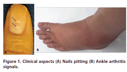
This case describes a caucasoid patient of the female sex, 32 years of age, without diagnosis. The patient referred red eyes when using a computer, with weak nails, with falls from some, left ankle swelling 6 months ago and sporadic pain in the Achilles tendon region for 12 months. On physical examination presented nails pitting, but without skin lesions, left ankle arthritis signals and pain on palpation in the Achilles tendon region (Figure 1).
Figure 1: Clinical aspects (A) Nails pitting (B) Ankle arthritis signals.
Ultrasound assessments
Written consent was obtained from patient to be evaluated at the rheumatology and ultrasonography service of the Catholic Pontifical University of Campinas. Sonographic evaluation was performed in the patient by a single rheumatologist with 12 years’ experience in exams. An ultrasound of the MyLab 50 equipment (Esaote S.p.A., São Paulo, Brazil) with 12 MHz high frequency linear probe was used to evaluate the eyes and a frequency of 15 to 18 MHZ for joint investigate, with power Doppler frequency of 6.6–8 MHz, Pulse Repetition Frequency that varied from 0.5 Hz to 1.0 MHz, and low filter. Ocular ultrasound evaluation was done with the eyes closed and with an important amount of gel and without pressing their structures and was asked the patient to move the eyes latero-medial, with a probe positioned longitudinally and transversely. All joints imaging were performed using gray scale (modo-B) and power Doppler techniques to detect morpho-structural changes and the presence of abnormal, following the OMERACT ultrasound guide for musculoskeletal evaluation [6].
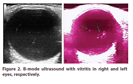
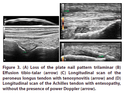
The ultrasound showed in eyes the following findings: confluent floating hypoecogenic images in vitreous humor more intensely to the left and sparse hypoecogenic images to the right, characterizing vitritis (Figure 2), in all of the distal interphalangicas evaluated, showed loss of the plate nail pattern trilaminar, in the left longitudinal scan of the tibio - talar with effusion, in the left longitudinal and transverse scan of the peroneus longus tendon presented tenosynovitis without power Doppler and in left longitudinal scan of the Achilles tendon showed an entesopathy, without the presence of power Doppler (Figure 3).
Discussion
The ultrasound eye assessment may be carried out either by the A-mode or by the B-mode, used by the ophthalmologist and radiologist, respectively, in which they are employed alone or simultaneously [1-3].
Modern ocular ultrasound allows for a non-invasive and dynamic approach to the various intraocular structures, which allows for the monitoring of these structures, especially when they cannot be observed due to the loss of transparency in the optical media (cornea, crystalline and vitreous body). The ultrasound also assesses the detachment of the posterior vitreous, macular edema, choroid detachments, thickening of the sclera and retinal detachment [4,5].
For the rheumatologist, an evaluation of the intraocular inflammatory processes, characterized by vitreous opacities, shall vitritis, often subclinical, dissociated from the clinical ophthalmological examination, is very important, because this condition can be associated at joint inflammatory process in activity. The specific findings for cases of intermediate uveitis are translated as confluent peripheral vitreous opacities (Snowballs) or Snowbanking, to B-mode is observed, multiple mobile structures, generally high mobility and low ecogenicity, can be confluent and occupy every vitreous cavity, we note important remission of these echoes when there is therapeutic effectiveness, so this method of image can be used to diagnose and monitor treatment [5].
Fifty to 80% of patients with PsO have concurrent nail lesions, which can lead to functional impairment, pain and discomfort, and decreased quality of life and general well-being. Despite its significant prevalence, nail manifestations are often neglected in daily clinical practice, probably due to a lack of recognition of its impact on patients or its relevance as an indicator of disease extension. Approximately 10%-37% of patients have skin and joint disease simultaneously, and 6%- 18% of patients have arthritis preceding psoriasis. This case report presented a patient with arthritis before skin lesion and ultrasound showed joints lesions findings [7,8].
In our rheumatological clinical practice, several cases of inflammatory arthropathy associated with uveitis evaluate, future studies will better signal the intensity of these findings of vitreous humor alterations, and also define this relationship in ocular changes and joint activity inflammatory process.
B-mode ocular ultrasound is an important adjunct to the clinical evaluation of a variety of eye diseases. When ophthalmoscopy is not possible, mainly due to opacification of the transparent environment (e.g. mature cataract or vitreous hemorrhage), or other structural eye interference that prevents evaluation, especially of the posterior chamber. Ultrasound is useful in eye examinations and portrays multiple conditions, such as inflammatory conditions, which may involve any structure of the eyeball [1,2].
Conclusion
Familiarity with the technique and eye anatomy encourages the use of this image method, like an important application and can guide the ophthalmologist and rheumatologist in the diagnosis of the uveitis mainly associated the arthritis to for a clear and safe diagnosis.
References
- De La Hoz Polo M, Torramilans Lluís A, Pozuelo Segura O et al. Ocular ultrasonography focused on the posterior eye segment: what radiologists should know. Insights. Imaging. 7(3), 351–364 (2016).
- Lorente-Ramos RM, Armán JA, Muñoz-Hernández A et al. US of the Eye Made Easy: A Comprehensive How-to Review with Ophthalmoscopic Correlation. RadioGraphics. 32(5), E175-E200 (2012).
- del Río LEC, González GAD, Ruiz MM et al. Characterization of cyclitic membranes by ultrabiomicroscopy in patients with pars planitis. J. Ophthal. Inflammation. Infection. (2020).
- Bedi DG, Gombos DS, Ng CS et al. Sonography of the Eye. Am. J. Roentgenol. 187(4), 1061–1072 (2006).
- Solarte CE, Shaikh A. Ultrasound techniques in ophthalmology. Ophthalmology. Pp, 137–149 (2007).
- Bruyn GA, Iagnocco A, Naredo E et al. OMERACT definitions for ultrasonographic pathology and elementary lesions of rheumatic disorders fifteen years on. J. Rheumatol. 46(10), 1388–1393 (2020).
- Mendonça JA. Differences of spectral Doppler in psoriatic arthritis and onychomycosis. Rev. Bras. Reumatol. 54(6), 490–493 (2014).
- Mendonça JA, Aydin SZ, D’Agostino MA. The use of ultrasonography in the diagnosis of nail disease among patients with psoriasis and psoriatic arthritis: a systematic review. Adv. Rheumatol. 59(41), 1–12 (2019).


