Clinical images - Imaging in Medicine (2018) Volume 10, Issue 6
18F-FDG uptake in inferior vena cava tumor thrombus
Patricia Barros Gouveia*Rua Maria Feliciana, N47 r/c direito, 4465-280, Sao Mamede Infesta, Porto, Portugal
- Corresponding Author:
- Patricia Barros Gouveia
Rua Maria Feliciana, N47 r/c direito
4465-280, Sao Mamede Infesta, Porto, Portugal
E-mail: patirigouveia@gmail.com
Abstract
Keywords
PET/CT, 18F-FDG, tumor thrombus, inferior vena cava, renal cell carcinoma
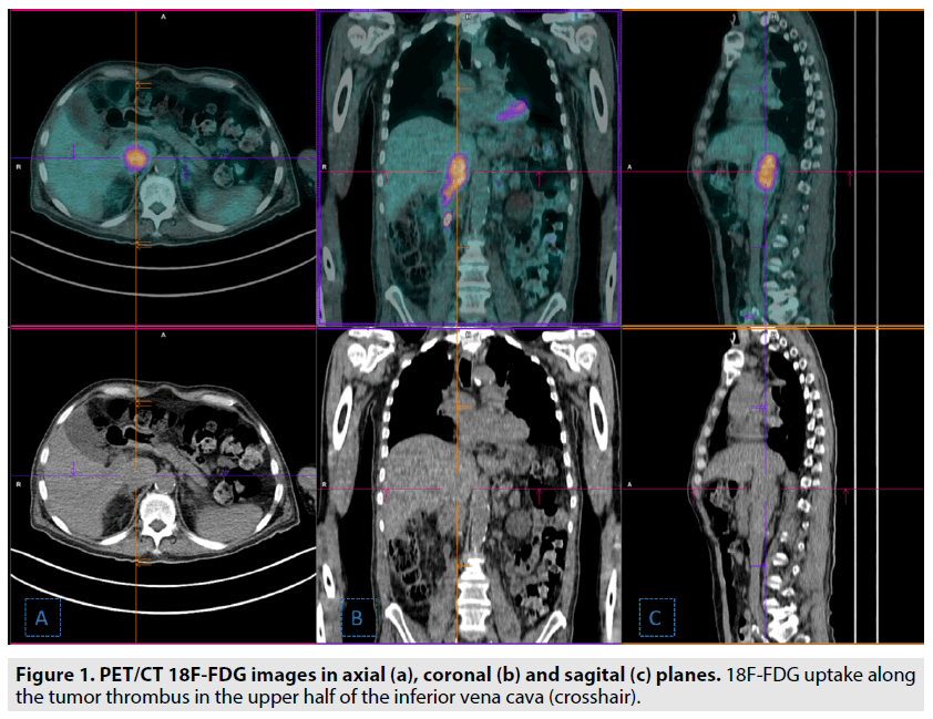
A 69 y old man with a past medical history of transglotic carcinoma was admitted to our institution with severe health status deterioration. The laboratory evaluation revealed a suppressed parathyroid hormone (PTH) level and hypercalcemia. He underwent a PET/ CT 18F-FDG scan to investigate a potential malignancy, which showed a linear tracer uptake along the upper half of the inferior vena cava (crosshair), suspicious for tumor thrombus. A Computed tomography angiography was performed and confirmed tumor thrombus in the inferior vena cava and revealed a rightsided kidney mass, compatible with renal cell carcinoma.
Intravascular tumor thrombus, defined as tumor extension into a vessel, is a rare complication of solid tumors [1,2]. Propagation of renal cell carcinoma into the inferior vena cava (IVC) has been reported in 4%-10% of patients [3,4]. This case illustrates the usefulness of PET/ CT 18F-FDG in identifying tumor thrombus and its contribution for an accurate staging (FIGURE 1).
Figure 1: PET/CT 18F-FDG images in axial (a), coronal (b) and sagital (c) planes. 18F-FDG uptake along the tumor thrombus in the upper half of the inferior vena cava (crosshair).
References
- Quencer KB, Friedman T, Sheth R et al. Tumor thrombus: incidence, imaging, prognosis and treatment. Cardiovasc. Diagn. Ther. 7: 165-177, (2017).
- Lai P, Bomanji JB, Mahmood S et al. Detection of tumour thrombus by 18F-FDG-PET/CT imaging. Eur. J. Cancer. Prev.16: 90-94, (2007).
- Giuliani L, Giberti C, Martorana G et al. Radical extensive surgery for renal cell carcinoma: long-term results and prognostic factors. J. Urol. 143: 468-473, (1990).
- Jungberg B, Stenling R, Osterdahl B et al. Vein invasion in renal cell carcinoma: impact on metastatic behavior and survival. J. Urol. 154: 1681-1684, (1995).


