Case Report - Imaging in Medicine (2018) Volume 10, Issue 2
Role of Ultrasonography in recognition of malignant potential of thyroid nodules on the basis of their internal composition
Sumanta Kumar Mandal & Sweta Singh*Department of Radiodiagnosis, Bankura Sammilani Medical College and Hospital, Bankura, West Bengal
- Corresponding Author:
- Sweta Singh
Department of Radiodiagnosis
Bankura Sammilani Medical College and Hospital, Bankura, West Bengal
E-mail: singhswetambbs@gmail.com
Abstract
Nodular thyroid is a common clinical entity. All patients were evaluated by grey scale USG and colour Doppler and then subjected to FNAC. Histopathology was done whenever required. The results were then compared. most of the benign nodules as well as the malignant nodules were predominantly solid or solid-cystic with predominant solid components.
Keywords
nodular thyroid, ultrasonography, malignant and benign tumour
Background and objectives
Nodular thyroid is a common clinical entity. The optimal diagnostic strategy to evaluate the nodule and differentiate it into benign and malignant, keeping in mind all the associated accompanying features is still a matter of debate. The present study was undertaken to evaluate the diagnostic efficacy of ultrasonography and fine needle aspiration cytology in differentiating benign and malignant thyroid nodules on the basis of composition (solid, cystic or mixed) [1-3].
Materials and methods
A prospective study was carried out in 65 patients of all age groups and both sexes attending the radiology department of Bankura sammilani medical college and hospital, Bankura, during the period of January 2018 to march 2018. These patients were referred to the department by Department of General Medicine, General Surgery and ENT.
Inclusion criteria-all patients with a palpable neck swelling clinically examined to be thyroidal in origin. Exclusion criteria-patients with diffuse neck swelling, ulcerative and fungating neck masses, moribund patients [4]. All patients thus included in the study, were evaluated by grey scale USG by the linear probe on HD7 Philips machine and colour Doppler and then subjected to FNAC after an informed consent was taken. Histopathology was done whenever required. The results were then compared.
Statistical analysis was conducted on a master chart created in MS Word and Excel sheet, evaluated by SPSS 17.0. Sensitivity, specificity, positive predictive value and negative predictive value were calculated to analyse the diagnostic accuracy of ultrasound and correlating with FNAC/HPE (Fine Needle Aspiration Cytology/ Histopathological Examination), as gold standard. For all statistical tests, p<0.005 was taken to indicate a significant difference.
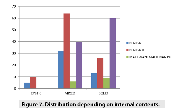
The majority of thyroid nodules were benign in nature [5]. The sensitivity and specificity of us diagnosing malignant lesions were 80% and 86%, respectively, out of 50 benign nodules, 64% were having mixed compositions. Out of 15 malignant nodules, 9 (60%) were found to be solid in composition. 23% benign nodules were solid compared to 60% solid malignant nodules.
Observations and results
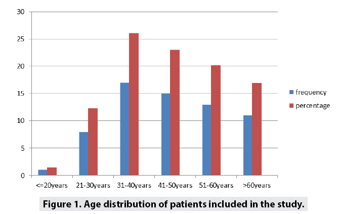
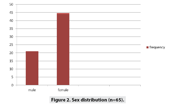
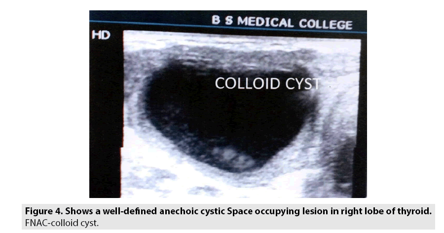
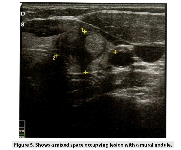
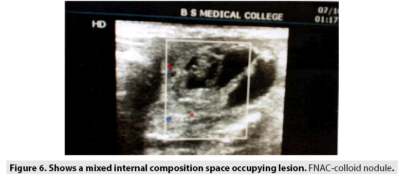
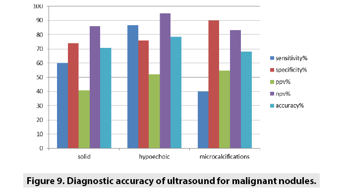
This study was conducted in the Department of Radio-diagnosis of Bankura Sammilani Medical College and Hospital, Bankura.65 patients were included in the study (Figures 1-9).
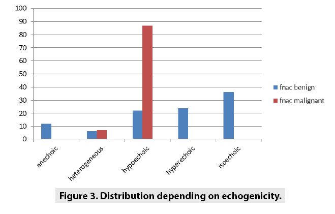
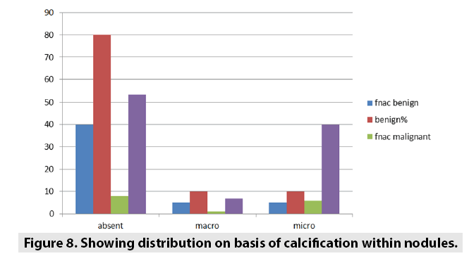
Interpretations and Conclusions in our study, most of the benign nodules as well as the malignant nodules were predominantly solid or solid-cystic with predominant solid components. Thus a predominantly solid component alone cannot be a useful criterion for the differentiation of malignant from benign nodules [6]. The positive predictive value of solid nodules is only 40% compared to 86%on negative predictive value. Out of 50 benign nodules, maximum were isoechoic to the normal glandular parenchyma. Hypoechoic appearance of malignant nodules is significant. Presence of micro calcifications in 40% malignant nodules as compared to 10% benign nodules is also significant (Tables 1-4) [7-11].
| Echogenicity | FNAC benign % | FNAC malignant % | P value |
|---|---|---|---|
| Anechoic | 12 | 0 | 0.322 |
| Heterogeneous | 6 | 6.7 | 1.0 |
| Hypoechoic | 22 | 86.7 | <0.001 |
| Hyperechoic | 24 | 0 | 0.05 |
| isoechoic | 36 | 6.7 | 0.049 |
Table 1: Distribution of nodules based on echogenicity.
| Internal contents | FNAC Benign Frequency |
FNAC Benign Percentage |
FNAC Malignant Frequency |
FNAC Malignant Percentage |
P Value |
|---|---|---|---|---|---|
| Cystic | 5 | 10 | 0 | 0 | 0.70 |
| Mixed | 32 | 64 | 6 | 40 | 0.06 |
| Solid | 13 | 26 | 9 | 60 | 0.015 |
Table 2: Distribution of thyroid nodules based on internal contents.
| Calcification | FNAC benign | % | FNAC malignant | % | P-value |
|---|---|---|---|---|---|
| Absent | 40 | 80 | 8 | 53.3 | 0.039 |
| Macro | 5 | 10 | 1 | 6.7 | 1.0 |
| Micro | 5 | 10 | 6 | 40 | 0.007 |
Table 3: Distribution of thyroid nodules based on calcification.
| USG features | Sensitivity % | Specificity% | Positive predictive value% | Negative predictive value% | Accuracy |
|---|---|---|---|---|---|
| Solid | 60 | 74 | 40.9 | 86 | 70.8 |
| Hypoechoic | 86.7 | 76 | 52 | 95 | 78.5 |
| Microcalcification | 40 | 90 | 54.5 | 83.3 | 68 |
Table 4: Diagnostic accuracy of Duplex ultrasound features for internal composition of malignant nodules.
Conclusion
In our present study, we found that, most of the benign nodules as well as the malignant nodules were predominantly solid or solid-cystic with predominant solid components. Thus a predominantly solid component alone cannot be a useful criterion for the differentiation of malignant from benign nodules. A combination of ultrasonographic features as well as pathological correlation is very necessary to increase the diagnostic accuracy and decrease the number of unnecessary invasive interventions in benign cases.
References
- Frates MC, Benson CB, Doubilet PM et al. Can color Doppler sonography aid in the prediction of malignancy of thyroid nodules? J. Ultrasound. Med. 22, 127-131 (2003)
- Enrico P, Rinaldo G, Antonio B et al. Risk of Malignancy in non-palpable thyroid nodules: Predictive value of ultrasound and color-Doppler features. J. Clin. Endocrinol. Metab. 87, 1941-1946 (2002).
- Iannuccilli JD, Cronan JJ, Monchik JM. Risk for malignancy of thyroid nodules as assessed by sonographic criteria: the need for biopsy. J. Ultrasound. Med. 23, 1455-1464 (2004)
- Gupta M, Gupta S, Gupta VB. Correlation of fine needle aspiration cytology with histopathology in the diagnosis of solitary thyroid nodule. J. Thyroid. Res. 10, 1-8 (2010)
- Iqbal M, Mehmood Z, Rasul S et al. Carcinoma thyroid in multi and uninodular goiter. J. Coll. Physicians. Surg. Pak. 20, 310-312 (2010).
- Delbridge L. Solitary thyroid nodule: Current management. ANZ. J. Surg. 76, 381-386 (2006).
- Mazzaferri EL. Management of a solitary thyroid nodule. N. Engl. J. Med. 328, 553-559 (1993).
- Kuru B, Gulcelik NE, Gulcelik MA et al. Predictive index for carcinoma of thyroid nodules and its integration with fine-needle aspiration cytology. Head. Neck. 31, 856-866 (2009).
- Kamran SC, Marqusee E, Kim MI et al. Thyroid nodule size and prediction of cancer. J. Clin. Endocrinol. Metab. 98, 564-570 (2013).
- Edino ST, Mohammed AZ, Ochicha O et al. Thyroid cancers in nodular goiters in Kano, Nigeria. Niger. J. Clin. Pract. 13, 298-300 (2010).
- Rago T, Di Coscio G, Basolo F et al. Combined clinical, thyroid ultrasound and cytological features help to predict thyroid malignancy in follicular and Hupsilonrthle cell thyroid lesions: Results from a series of 505 consecutive patients. 66, 13-20 (2007).
