Clinical images - Imaging in Medicine (2020) Volume 12, Issue 1
Nasal NK/T-cell lymphoma: Clinical features and dermoscopic appearances
Jihane Ziani*, Mounia Bennani, Zakia Douhi, Sara Elloudi, Hanane Baybay & Fatima Zahra MernissiDepartment of Dermatology, Hassan II hospital university, Fez, Morocco
- Corresponding Author:
- Jihane Ziani
Department of Dermatology
Hassan II hospital university, Fez, Morocco
E-mail: dr.zianijihane@gmail.com
Abstract
Keywords
NK/T-cell lymphoma ■ dermoscopy appearance
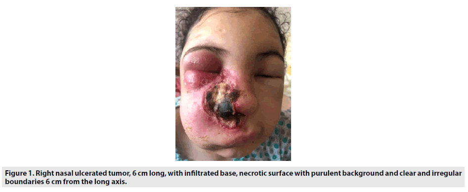
CA 18y old female without family history, who presented 6 months before, a right nasal swelling with the notion of right nasal obstruction evolving towards an ulcerated tumor whose biopsy returned in favor of a nasal NK lymphoma. Physical examination showed a right nasal destructive ulcerated tumor, with an infiltrated base, with a necrotic surface with a purulent background and with clear and irregular boundaries making 6cm of the long axis (FIGURE 1). In dermoscopy appearance we had especially an aspect in gaps with separations by whitish septa (FIGURE 2).
Nasal NK/T-cell lymphoma occurs in adult males generally in the fifth decade. The male/female sex ratio is 3:1 and the median age at diagnosis is 50-60 years. The clinical presentation is dominated by local signs such as unilateral nasal obstruction, purulent and/ or ribbed rhinorrhea, recurrent epistaxis and chronic sinusitis.


