Case Report - Imaging in Medicine (2022) Volume 14, Issue 7
Middle Turbinate Mucocele: An Unsual Location Mucocele Du Cornet Moyen: Une
Chiboub D*, Romdhane N, Belaid H, Ouerghi A, Nefzaoui S, Hariga FI, Mbarek Ch
ENT Department of Habib Thameur Hospital
Received: 29-Jan-2022, Manuscript No. FMIM-22-52849; Editor assigned: 17-Jun-2022, PreQC No. FMIM-22-52849(PQ); Reviewed: 1-July-2022, QC No. FMIM-22-52849; Revised: 08-July-2022, Manuscript No. FMIM-22-52849(R); Published: 15-July-2022, DOI: 10.37532/1755-5191.2022.14(7).01-04
Abstract
Introduction: Nasal mucoceles are rare. The symptoms depend on the extension of lesion. They are especially related to nasal obstruction. It causes progressive distension inducing organ compression and bone erosion. Most commonly seen in the frontoethmoidal area, it may also develop in abnormally aerated bones, such as middle turbinate, clinoid process and pterygoid process. Purpose: To report a rare location of a nasal mucocele and describe its different clinical and paraclinical aspects. Observation: We report a case of a 55-year old female with no medical history, who was complaining about visual acuity reduced on the left eye, which appeared during the past ten months. Endoscopic nasal examination revealed a formation of the middle left turbinate. CT scan (computed tomography scan) and MRI (magnetic resonance imaging) revealed the presence of a cystic lesion in the middle turbinate. This lesion was well-defined and homogeneous. CT scan revealed an erosion of the lamina papyracea and a compression of the internal rectus muscle. The MRI showed the orbital extension. The patient was operated with an endoscopic approach. We evacuated the content of the mucocele. The evolution was favorable with no recurrence during 2 years. Conclusion: The mucoceles remain asymptomatic for a long time, which can retard the diagnosis. Its peculiarity lies more in the diagnosis than in the management as most of these can be completely resected or marsupialized endoscopically. The imaging is indicated before the surgical treatment.
Keywords
Mucocele • middle turbinate • computed tomography scan • magnetic resonance imaging
Introduction
The mucocele is a benign pseudocyst. It causes progressive distension inducing organ compression and bone erosion [1]. Most commonly seen in the frontoethmoidal area, these mucoceles develop as a result of obstruction of the normal sinus drainage tract. Rarely, it may also develop in abnormally aerated bones, such as middle turbinate, clinoid process and pterygoid process. Obstruction of the concha bullosa can rarely lead to formation of a mucocele which may be secondarily infected forming a mucopyocele. We report the case of an adult operated for a middle turbinate formation related to mucocele.
■Observation
We report a case of a 55-year old patient, with no surgical or traumatic past history, complaining of visual acuity reduced on the left eye, which appeared during the past ten months. There is no headache neither rhinologic symptoms. On examination, no face swelling or lymph nodes were found.
Nasal endoscopy revealed an enlarged middle turbinate occupying almost the entire left nasal cavity, pushing the nasal septum to the right side. The right nasal cavity and the nasopharynx were normal.
Specialized ophtalmological examination found no proptosis and the visual acuity was 3/10 on the left eye and 7/10 on the right eye.
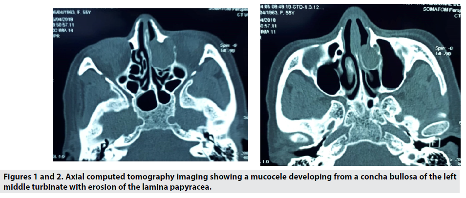
CT scan revealed a well circumscribed expansile mass of the middle turbinate with an effect on the lamina papyracea (Figures 1 and 2). This mass was isodense with a compress of the internal rectus muscle.
Figures 1 and 2 Axial computed tomography imaging showing a mucocele developing from a concha bullosa of the left middle turbinate with erosion of the lamina papyracea.
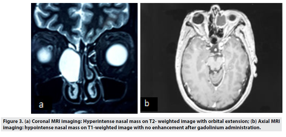
MRI was indicated because of the orbital extension. The mucocele appeared as homogeneous mass in low signal on T1- weighted images, high signal on T2-weighted images (Figure 3) and with no enhancement after gadolinium administration. Orbital content was repulsed and optic nerve was untouched.
The patient had an endoscopic treatment. A surgical marsupialization of mucocele and turbinoplasty were performed. The evolution was favorable with no reccurence during 2 years. Visual acuity in the left eye was improved significantly up to 5/10.
Discussion
A mucocele is a cystic mass filled with mucus and lined by respiratory epithelium. It can cause an erosion of the bone or a compression of adjacent anatomical structures [2,3].
Turbinate location is unusual. They are commonly found in the middle turbinate because of its pneumatization [2].
Concha bullosa is the most frequently encountered anatomical variation of the lateral nasal wall. Mucoceles develop as a result of impaired drainage of the mucus, which can either be caused by alteration in its viscosity or composition as seen in cystic fibrosis or by mechanical obstruction.
Mucoceles are mostly painless and asymptomatic swellings that have a relatively rapid onset and fluctuate in size [4-6]. Their enlargement may cause clinical symptoms (nasal obstruction, rhinorrhea, facial pain or ophtalmic complications). The lesion can compress the orbit inducing a raised intraocular pressure, diplopia, or proptosis [7]. Mucoceles of the middle turbinate have been associated with nasal obstruction, nasal discharge, diplopia, exophthalmos and chronic sinusitis symptoms owing to the blockage of ethmoidal infundibulum [8].
In our case, the patient complained about visual acuity reduction without any rhinological symptoms.
Physical examination presents with ophtalmic abnormality like visual disturbance or impaired ocular mobility. Nasal endoscopy reveals an hypertrophy of the middle turbinate.
CT scan is the most helpfull investigation of mucoceles. They appear as a homogeneous, welldefined, non-enhancing, expansile cystic lesion in the middle turbinate [3,5].
In some cases, CT scan inform about causes of mucoceles as adhesions, concha bullosa, septum deviation, paradoxical middle turbinate. This exam allows also defining anatomic variants prior to surgery [3,9].
MRI allows to differentiate between mucocele or other tumors and to analyze orbital and intracranial extension in complement of the CT scan.
In our study, MRI was necessary to evaluate orbital extension. The compression of the internal rectus muscle and the occular globe was found in MRI but with no diplopia in ophtalmological examination [3,10,11].
Mucocele may have different signals on MRI depending on its composition [12]. A low intense T1 and a high intense T2 is usually found [3].
The treatment is based on an endoscopic surgery as soon as possible for symptomatic mucocele [13-15].
This procedure had a low recurrence rate [16]. A middle turbinoplasty can be also performed.
A special care must be taken when removing mucocele walls adjacent to the orbit or skull base.
Conclusion
The clinical features of middle turbinate mucocele are diverse. It represents a diagnostic challenge to surgeons both in terms of symptoms and risk of complications. Its peculiarity lies more in the diagnosis than in the management as most of these can be completely resected or marsupialized endoscopically.
References
- Erdogan BA, Unlu N, Aydin S et al. Frontal Mucocele Extended Orbita and Endoscopic Marsupialization Technique. J Craniofac Surg. 29, e408‑9 (2018).
- Lee YW, Kim YM. Mucocoele of the inferior turbinate: a case report. Br J Oral Maxillofac Surg. 54, 1121‑2 (2016).
- Marrakchi J, Nefzaoui S, Chiboub D. Imaging of paranasal sinus mucoceles. Otolaryngol Open J. 2, 94–100 (2016).
- Pinto JA, Cintra PP, De Marqui ACS et al. Middle turbinate mucopyocele: a case report. Rev Bras Otorrinolaringol. 71, 378-81 (2005).
- Cinar U, Ozgur Y, Uslu B et al. Pyocele of the middle turbinate: a case report. Kulak Burun Bogaz Ihtis Derg. 12, 35-8 (2004).
- Bahadir O, Imamoglu M, Bektas D. Massive concha bullosa pyocele with orbital extention. Auris Nasus Larynx. 33, 195-8 (2006).
- Kim JS, Kim EJ, Kwon SH. An ethmoid mucocele causing diplopia: A case report. Medicine (Baltimore). 96, e9353 (2017).
- Gupta N, Kalia M, Dass A et al. Mucocele of the middle turbinate: A rare entity. Roman J Rhinol. 10, 59-62 (2020).
- Xu Y, Tao Z, Zhan H et al. Massive concha bullosa pyocele with orbital extension-a case report and review of the literature. J Clin Otorhinolaryngol Head Neck Surg. 21, 1085‑6 (2007).
- Lee TJ, Li SP, Fu CH. Extensive paranasal sinus mucoceles: A 15-year review of 82 cases. Am J Otolaryngol. 30, 234-238 (2009).
- Kim YS, Kim K, Lee JG et al. Paranasal sinus mucoceles with ophthalmologic manifestations: A 17-year review of 96 cases. Am J Rhinol Allergy. 25, 272-275 (2011).
- Haloi AK, Ditchfield M, Maixner W. Mucocele of the sphenoid sinus. Pediatr Radiol. 36, 987-990 (2006).
- Ketenci I, İlhan Şahin M, Vural A. Mucopyocele of the Concha Bullosa: A Report of Two Cases. Erciyes Med J. 35, 157-60 (2013).
- Lee. Concha bullosa mucocele with orbital invasion and secondary frontal sinusitis: a case report. BMC Res Notes. 6, 501 (2013).
- Devars du Mayne M, Moya-Plana A, Malinvaud D et al. Sinus mucocele: Natural history and long-term recurrence rate. Eur Ann Otorhinolaryngol Head Neck Dis. 129, 125-30 (2012).
- Mora-Horna ER, Lopez VG, Anaya-Alaminos R et al. Optic neuropathy secondary to a sphenoid-ethmoidal mucocele: case report. Arch Soc Eps Oftalmol. 90, 582–4 (2015).
Indexed at, Google Scholar, Crossref
Indexed at, Google Scholar, Crossref
Indexed at, Google Scholar, Crossref
Indexed at, Google Scholar, Crossref
Indexed at, Google Scholar, Crossref
Indexed at, Google Scholar, Crossref
Indexed at, Google Scholar, Crossref
Indexed at, Google Scholar, Crossref
Indexed at, Google Scholar, Crossref


