Clinical images - Imaging in Medicine (2018) Volume 10, Issue 3
Beaver tail liver in evans syndrome due to systemic lupus erythematosus
Al Attia HM1*, Imran Khan2& Khateeb KAL21Private Practice,Abu Dhabi, United Arab Emirates
2Department of Radiology, Private practice, Universal Hospitals, Abu Dhabi, United Arab Emirates
Abstract
Keywords
SLE ▪ beaver tail liver ▪ evans syndrome ▪ thrombocytopenia
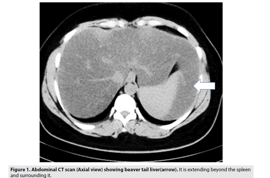
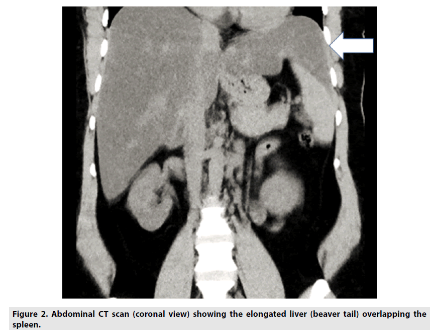
A 33 year old female diagnosed as Evan’s syndrome due to systemic lupus erythematosus (SLE), and lupus nephritis was further investigated for clinically enlarged liver and spleen [1]. The CT abdomen showed in addition to a mild hepato-splenomegaly, the presence of significant beaver tail liver (FIGURES 1 &2). Though this incidental finding is of a benign nature it yet has some clinical implications particularly in a patient like this one, who has thrombocytopenia (platelets of 86.000 mm3), and hemorrhage is a potential complication. In case of trauma or injury to left side of the abdomen, which classically affects the spleen, this liver elongation may be affected and can be mistaken for splenic trauma or hemorrhage. Furthermore, the associations of this liver variant with other conditions are rather rare in the literature, so its coexistence here with a case of SLE related Evan’s syndrome has some academic orientation as well.


