Case Report - Imaging in Medicine (2017) Volume 9, Issue 5
An unusual sonographic appearance of a dermoid cyst in a young patient
Moawia Bushra Gameraddin1*, Sultan Alshoabi1 & Yousif Alarkani21Faculty of Applied sciences, Department of Diagnostic Radiologic Technology, Taibah University, Saudi Arabia
2Department of Radiology, King Fahad Hospital, Saudi Arabia
- Corresponding Author:
- Moawia Bushra
Gameraddin
Faculty of Applied sciences
Department of Diagnostic Radiologic Technology
Taibah University, Saudi Arabia
E-mail: m.bushra@yahoo.com
Abstract
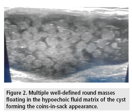
A dermoid cyst is a rare benign cystic teratoma. On ultrasound, it has variable sonographic features since it is usually filled with an admixture of ectoderm, mesoderm, and endoderm origin contents. Because of the lumen of the dermoid cyst is usually filled with an admixture of ectoderm, mesoderm and endoderm origin contents, it appears as different areas of echogenicity. The coalescence of the hyperechoic fat into globules within the hypoechoic fluid matrix forming an unusual appearance of ''Coins-in-Sack'' on ultrasonography. This is virtually typical for a dermoid cyst. In this case, we suggested the term "Coins-in-Sack" appearance as an accurate and descriptive diagnostic sonographic feature of this an uncommon para-sacral dermoid cyst.
Keywords
unusual ▪ sonographic ▪ appearance ▪ dermoid ▪ cyst ▪ coins in-sack
Introduction
A dermoid cyst is a benign cystic teratoma that is a congenital and/or developmental in origin and usually present in young adults frequently located in the midline of the human body. Most dermoid cysts are considered uncommon congenital non-neoplastic lesions. They are composed of epidermal elements and dermal derivatives surrounded with a capsule [1].
A dermoid cyst is usually a well-defined, unilocular and thin wall cyst that may show the characteristic "Sac-of-marbles" appearance on ultrasonography due to the coalescence of fat into globules of fat floating in the fluid matrix within the lumen of the cyst [2].
Dermoid cysts usually originate from the outer germ layer ectoderm due to the abnormal closure of the ectodermal tube but it may have contents from the three embryonic germ layers' ectoderm, mesoderm, and endoderm. Infection of dermoid cyst can occur especially when it is misdiagnosed as an abscess and undergone to surgical intervention. Despite the uncommon occurrence of a dermoid cyst in the retro-rectal region, comprehensive study of the cause, presentation, medical imaging and management of these lesions is very important as the incorrect diagnosis of dermoid cyst can lead to incorrect management that can result in the reverse outcome and worse health problems [3].
Case Study
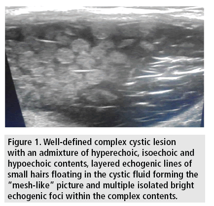
A 21 years old male was complaining of a painless swelling at the upper inner quadrant of his right buttock for three months. The provisional diagnosis was a gluteal abscess but there were no tenderness and no fever, so, he was referred for ultrasound imaging. The ultrasound scanning was performed using 7.5 MHz transducer and revealed a well-defined complex heterogeneous echogenicity cystic lesion of approximately 6*7 cm in maximal transverse dimensions that contains an admixture contents of mixed echogenicity with/without acoustic shadows including multiple well-defined hyperechoic balls forming the Coins in Sack appearance and multiple echogenic linear contents in a hypoechoic fluid matrix. A biopsy was done and histopathology results revealed no malignant cells in this cystic lesion. These findings are consistent with a dermoid cyst. Informed consent was obtained from the patient to participate in the case study (FIGURES 1 and 2).
Discussion
A dermoid cyst is an abnormal growth that frequently affects the ovaries [4], but additional locations include the cranium [5], parotid gland and the floor of the mouth [6,7]. They approximately represent 20% of intradural tumors in children specifically in the 1st year of life [8]. They are rarely involving the buttock. Histologically, the dermoid cyst may contain any tissue, including hair, tooth, cartilage, fluid, skin glands and gastrointestinal tissue [9]. Since these cysts contain variable components of tissues, the sonographic display exhibit different echogenicity, and different appearances. These cysts contain mostly sebaceous fluid which appears on ultrasound as a hypoechoic cyst with echogenic components that represent a mixture of hair, more solid sebaceous material and strands of fibrous tissue. These components may exhibit echogenic lines [10,11].
In the current case, the patient presents with a painless lump in his buttock for three months' period, particularly due to the patient’s medical history, clinical examination and laboratory analysis of the patient's blood. The diagnosis of an abscess could be ruled out in comparison with Sung, who reported that all patients with gluteal abscess were present with raised inflammatory process [12]. The diagnosis of a malignant mass was excluded in this case since the histopathological findings revealed no malignant changes. In a previous study, incidence of malignant transformation of dermoid cysts was estimated to be 0.17-2% [13]. This confirmed the benignancy of demoid cysts.
In this case study, there were multiple uniformly hyperechoic nodules and multiple layered bright linear echoes and isolated punctate echogenic foci within the contents of the lesion. These findings are strong sonographic characteristics of a dermoid cyst according to Ameye et al. [14], who reported specific diagnosis of dermoid cysts using ultrasound. They reported that mixed echogenicity of dermoid cysts representing the different components of fat, bone and fluid. However, the previous sonographic features can be confidentially diagnosed as a dermoid cyst.
The sensitivity and specificity of ultrasound imaging in diagnosing dermoid and epidermoid cysts were approximately 80.0% and 95.4%, respectively [15]. Therefore, it is capable to differentiate dermoid cysts from other softtissue masses.
Finally, this case describes rare features of a dermoid cyst using ultrasonography which may be used to predict specific type of other teratomas. However, magnetic resonance imaging (MRI) is recommended for further demonstration of soft tissue contents in the dermoid cyst.
Conclusion
In this case, we have described a rare sonographic appearance of a type of dermoid cysts. Coins-in-Sack appearance is a very rare sonographic feature that we have suggested in this case. It describes and characterizes this rare para sacral dermoid cyst. It might be considered as a pathognomonic sonographic sign for the diagnosis of dermoid cyst.
Financial Support and Sponsorship
Nil
Conflicts of Interest
There are no conflicts of interest.
References
- Pant I, Joshi SC. Cerebellar intra-axial dermoid cyst: A case of unusual location. Childs. Nerv. Syst. 24, 157-159 (2008).
- Sanal HT. Ultrasound findings of dermoid cysts located in neck region: Sack-of-Marbles sign. KBB-Forum. 7 (2008).
- Hassan I, Wietfeldt ED. Presacral tumors: Diagnosis and management. Clin. Colon. Rectal. Surg. 22, 84-93 (2009).
- Longo DL, Fauci AS, Kasper DL et al. Harrison's principles of internal medicine, 18e. New York, NY: McGraw-Hill. (2012).
- Majed M, Nejat F, Khashab M. Congenital dermoid cysts of the anterior fontanel. Indian. J. Plast. Surg. 41, 238-240 (2008).
- Tas A, Yagiz R, Altaner S et al. Dermoid cyst of the parotid gland: first pediatric case. Int. J. Pediatr. Otorhinolaryngol. 74, 216-217 (2010).
- Vargas Fernandez JL, Lorenzo Rojas J, Aneiros Fernandez J et al. Dermoid cyst of the floor of the mouth. Acta. Otorrinolaringol. Esp. 58, 31-33 (2007).
- Kwinta B, Adamek D, Moskała M et al. Tumours and tumour-like lesions of the spinal canal and spine. A review of 185 consecutive cases with more detailed close-up on some chosen pathologies. Pol. J. Pathol. 62, 50-59 (2011).
- Aster JC, Abbas AK, Robbins SL et al. Robbins basic pathology. Ninth edition. Philadelphia, PA: Elsevier Saunders. (2013).
- Smorgick N, Maymon R. Assessment of adnexal masses using ultrasound: A practical review. Int. J. Womens. Health. 6, 857-863 (2014).
- Garner JP, Meiring PD, Ravi K et al. Psoas abscess-not as rare as we think? Colorectal. Dis. 9, 269-274 (2007).
- Jung S. Ultrasonography of ovarian masses using a pattern recognition approach. Ultrasonography. 34, 173-182 (2015).
- O’Neill K, Cooper AR. The approach to ovarian dermoids in adolescents and young women. J. Pediatr. Adolesc. Gynecol. 24, 176-180 (2011).
- Ameye L, Timmerman D, Valentin L, et al. Ultrasound. Obstet. Gynecol. 40, 582-91 (2012).
- Hung EH, Griffith JF, Hung AW et al. Ultrasound of musculoskeletal soft-tissue tumors superficial to the investing fascia. Am. J. Roentgenol. 202, W532-W540 (2014).


