Clinical images - Imaging in Medicine (2018) Volume 10, Issue 5
Urachal Cyst Presenting As A Pelvic Mass
Sindhu Kumar*Department of Radiology, University of Virginia Medical Center, Virginia 22901, USA
- Corresponding Author:
- Sindhu Kumar
Department of Radiology, University of Virginia Medical Center
Virginia 22901, USA, E-mail: sindhucumar@gmail.com
Abstract
Keywords
computed tomography
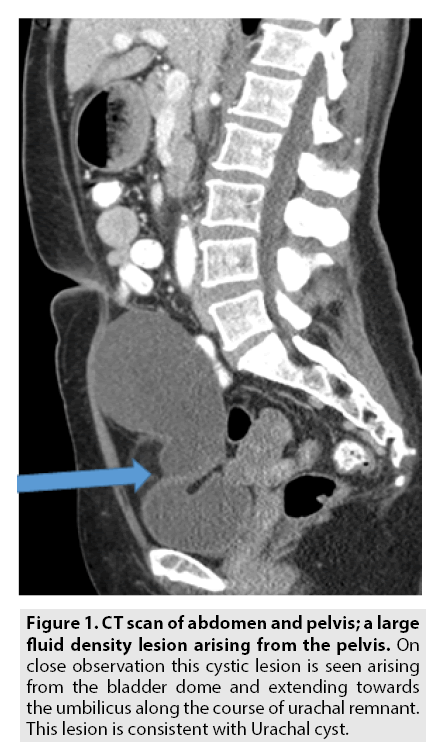
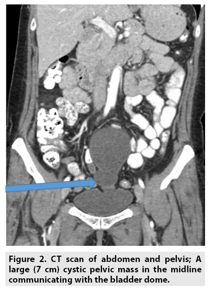
A urachal cyst occurs in the urachal remnant between the umbilicus and bladder [1]. It can present as an extraperitoneal mass in the umbilical region. It is characterized by abdominal pain, and fever if infected. It may rupture, leading to peritonitis or it may drain through the umbilicus. Urachal cysts are usually silent clinically until infection, calculi or adenocarcinoma develops. This patient was asymptomatic who presented with newly discovered pelvic mass (FIGURES 1 and 2).


