Research Article - Imaging in Medicine (2017) Volume 9, Issue 3
Thyroid shield during brain CT scan: dose reduction and image quality evaluation
Abuzaid MM1*, Elshami W1, Haneef C2 & Alyafei S21University of Sharjah, Medical Diagnostic Imaging Department, UAE
2Fatima College of Health Sciences, Radiography and Medical Imaging Department, UAE
- Corresponding Author:
- Abuzaid MM
University of Sharjah
Medical Diagnostic Imaging Department, UAE
E-mail: mabdelfatah@Sharjah.ac.ae
Abstract
Keywords
thyroid gland ▪ thermoluminescent dosimeter ▪ CT radiation dose ▪ radiation dose reduction ▪ MDCT
Introduction
Radiation doses during CT examinations are known to be relatively high and provide over 35- 40% of the collective radiation received from diagnostic imaging procedures [1]. The extensive use of CT and raised concerns about growing radiation exposure is empathizing the need for appropriate strategies to optimize and thereby, if possible, reduce radiation dose due to CT. Cancer risks associated with medical diagnostic radiation estimated that 29,000 future cancers (approximately 2% of the cancers diagnosed yearly in the United States could be related to (CT) [2].
Radiation received by organs in the primary beam can be clinically justified. However, the dose received by organs outside the primary beam cannot be justified when the organs are radiosensitive. Thyroid, eyes, breasts and gonads receive a greater proportion of radiation due to increased scatter and the lack of other overlying tissues to partially absorb some of the dose [3]. Reducing scatter doses without affecting the image quality must be implemented, solutions to reduce these radiation exposures like tube current, collimation, pitch, gantry rotation time and shielding [4].
Hein et al. reported that the use of a shield for protection of eye lenses in paranasal sinus CT is a suitable and efficient means of reducing radiation to the lens of the eye during routine cranial CT by 40-50% depending on the thickness of the shielding [5]. Hopper described the use of bismuth radioprotective garments to shield eyes, thyroid, and breast diagnostic CT while preserving the overall quality of the diagnostic image; 57% reduction in breast doses during thoracic CT with the use of bismuth radioprotective latex [3].
Scientists have proved a definite link between thyroid cancer and radiation but have struggled to calculate the risks from low doses such as those in medical exposures [6,7]. The thyroid gland is a major radiosensitive organ that exposed to scatter radiation during brain CT scan. Therefore, in addition to the other dose reduction measures taken in CT head exams, shielding of the thyroid has to be seriously considered.
Thyroid and breast shielding during CT head were reduced by an average 45% a 76%, respectively, in 110 patients undergoing routine head CT [8]. McLaughlin et al. recommended the use of thyroid shield for all CT scanning of the chest with an approximate 57% and 18% reduction in the thyroid and eye doses respectively [9]. Williams et al. reported that the effectiveness of shield varied with scanning technique used and that the thyroid dose due to scattered radiation was significantly reduced 46- 58% and 36-44% at the surface of the thyroid and within the thyroid tissue at 1 cm depth respectively after the use of a lead thyroid shield [7]. Mazonakis et al. [6] concluded that the scattered dose to the thyroid from the head CT examinations varied from 0.6 mGy to 8.7b mGy and the primary irradiation of the thyroid gland during CT of the neck caused by an absorbed dose range of 15.2-52.0 mGy.
Materials and methods
The aims of this study were to investigate the dose reduction to the thyroid gland during routine brain CT examinations. Also, the study compares the quality of the images before and after applying the thyroid shields.
An experimental study was done utilizing CT machine multi-slice 16, Toshiba Alexion and standard head attenuating phantom. Radiation dose to the thyroid during the scan recorded by TLD chips (Type GR 100 Lif MG, Ti: 3.2*3.2*0.89 mm). Since the rotation from the CT is multi-directional, a circumferential thyroid shielding (0.5 mm Pb equivalent) used to protect the thyroid in the experiment.
As a preparation for the experiment, the TLD chips annealed to erase any previous radiation present. A part of the annual quality control done to the CT machine before performing an experiment to make sure a consistent experiment. Quality control is done for laser accuracy, table travel, CT number accuracy, slice thickness and dose measurements. The thyroid shielding was also checked for any damages using fluoroscopy before the experiment.
For the experiment, nine TLDs enclosed in numbered plastic packets placed on the thyroid area of the phantom to measure the scattered radiation to the thyroid, without applying the shield (unshielded dose). The TLD sites were marked to ensure reproducibility for the next TLD set. The experiment repeated with the thyroid shielding implemented with a different set TLDs placed on the marked sites to measure the scattered dose after use of shielding (shielded dose). The scan parameters that were used during the scan was, 120 kVp, 180 mA, 5 mm Slice thickness, rotation time 2. The TLD chips read using HARSHAW QS 5500 TLD.
For the quality of the CT images with and without the thyroid shielding, two radiologists experienced in the interpretation of CT images did a qualitative analysis of images. A meeting between the two radiologists organized to discuss the controversies between their results and to come out with one decision. The images were assessed for parameters like grayscale (contrast), noise, soft tissue sharpness, bone detail and overall image quality using five-point images scale 1: not acceptable, 2: substandard, 3: acceptable, 4: above average and 5: excellent.
Results
Out of nine TLD used, four were eliminated due to discrepancies in the calefaction. TABLE 1 shows the results of the scattered dose evaluation to study the effectiveness of thyroid shielding in brain CT scans. The data shows the TLD readings with (shielded dose) and without (unshielded dose) thyroid shielding at the same points in the thyroid area. The calculated ratios of the unshielded and shielded doses along with percentage reductions at individual points are shown in the table. The dose reductions ranged from about 40-60%. The highest ratio of the dose without shielding to the dose with shielding [(W/o)/W] was 2.7 and the maximum reduction in the thyroid surface dose was 62.9%. The results show that there is a significant decrease in the entrance skin dose to the thyroid after the use of the shielding.
| TLD No. | Without shield | With shield | Calculations | |||
|---|---|---|---|---|---|---|
| Reading | Reading | Reading | Reading | Ratio | % | |
| (cGy) | (mGy) | (cGy) | (mGy) | (W/o) IW | Reduction | |
| 1 | 0.0257 | 0.26 | 0.0123 | 0.12 | 2.09 | 52.1% |
| 2 | 0.0173 | 0.17 | 0.0104 | 0.10 | 1.66 | 39.9% |
| 3 | 0.0133 | 0.13 | 0.0122 | 0.12 | 1.09 | 8.3% |
| 4 | 0.0172 | 0.17 | 0.0103 | 0.10 | 1.67 | 40.1% |
| 5 | 0.0248 | 0.25 | 0.0092 | 0.09 | 2.70 | 62.9% |
Table 1: Thyroid doses with and without thyroid shielding.
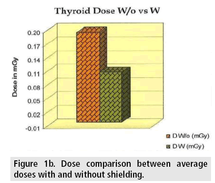
The mean doses to the thyroid area with and without the use of thyroid shielding along with the standard deviations are presented in TABLE 2. The average entrance dose to the thyroid was 0.20 mGy without the utilization of the shielding while the use of shielding reduced the mean dose to 0.11 mGy. The standard deviations were 0.05 and 0.01 for the unshielded and shielded doses respectively. The mean percentage dose reduction obtained was 44.7%. The dose reduction was also significant showing p-value of 0.027 (2 tailed).
| Without shield | With Shield | Dose Calculation | ||||||||
|---|---|---|---|---|---|---|---|---|---|---|
| Dose | +/- | n | Dose | +/- | n | D R | D R % | T | df | P (tailed) |
| mGy | SD | mGy | SD | |||||||
| 0.20 | 0.05 | 5 | 0.11 | 0.01 | 5 | 0.09 | 44.7 | 3.3 | 4 | 0.027 |
Table 2: The mean doses with and without thyroid shielding.
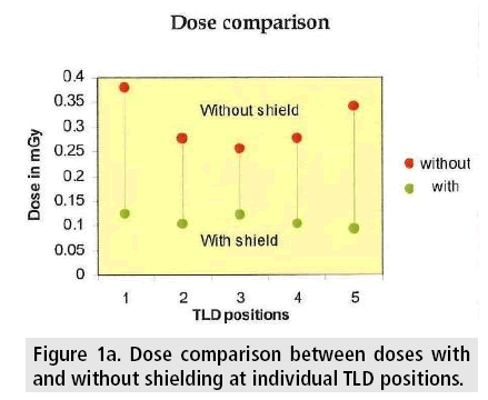
FIGURES 1a-b illustrates the dose reduction achieved with the use of thyroid shielding. FIGURE 1a shows the dose reduction at individual points of the TLD placement and FIGURE 1b illustrates the comparison between the average dose without the shielding and the average dose with shielding. The Y axis in the graphs show the doses in mGy units, and the X axis in FIGURE 1a shows the individual TLD positions.
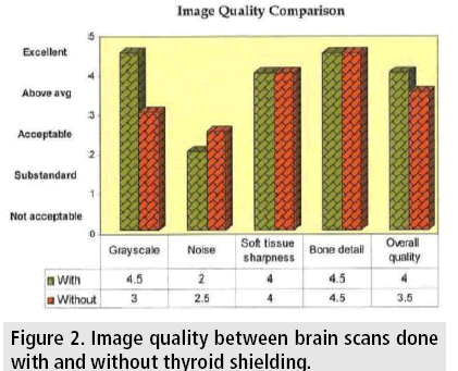
FIGURE 2 along with the data table illustrates the results of the study comparing the image quality between brain scans done with and without thyroid shielding. The overall image quality and the grayscale factor was higher for the brain scan done with the use of thyroid shielding whereas the grades received for soft tissue sharpness (4) and bone detail (4.5) were the same for both the scans (with and without thyroid shielding). For the noise factor, the scan done without the shielding showed a better quality. In sum, the result indicates that the use of shielding could cause no degradation to the image quality.
Figure 2: Image quality between brain scans done with and without thyroid shielding.
Discussion
The experimental result presented that there is a significant reduction in the entrance skin dose to the thyroid after the use of the shielding. The highest dose reduction achieved was 62%, and the lowest was 8.3%. The average decrease in the thyroid dose was 44.7%, and the radiation dose was reduced by a mean factor of 1.81 mGy. This indicates the importance of the use of thyroid shielding in patients undergoing brain CT scan especially pediatric patients who are much more sensitive to the radiation [10,11]. A low 8%, as well as a high 60% the reduction, was demonstrated at different points in the thyroid area. These differences could be due to the random distribution of the internal scatter radiation within the body part, giving varying dose reductions at different parts of the region. However, considering these readings would still have shown a 20% mean dose reduction and a low radiation reduction factor of 1.27 mGy. Previous studies reported a 46-58% dose reduction similar to the present findings. However, the highest unshielded dose to the thyroid reported being 0.69 mGy, which is different from the current study where the maximum dose recorded without shielding, is 0.26 mGy. This difference in the dose could be attributed to the difference in the scanning technique especially the kVp and mA settings [4,11,12]. The change in the scanning parameters playing the major role in contributing to the dose reduction which could be used as an indication to recommend the use of these parameters for routine brain CT scans. The image quality results (FIGURE 2) show that there is no degradation of the image quality due to the use of thyroid shielding. The scans done after applying the thyroid shielding showed better quality regarding grayscale and the overall image quality factor than those done without the shielding. One possible reason for this increased image quality could be the absorption of the scatter radiation by the shielding, considering the general fact that scatter radiation usually degrades the picture quality. The image detail factors like soft tissue sharpness and bone detail showed absolutely no difference whether it was a scan done with the shielding or without the shielding. In the scans done with the thyroid shield, the noise factor was graded substandard while that in the unshielded scan was acceptable. Another unique point to be added is that the images were given for quality assessment as they were obtained from the scanner without any manipulations done. The application of thyroid shielding at the short neck patient can be difficult. In these cases, sometimes applying thyroid shielding can even be risky causing artifacts in the image. One solution to this would be to implement the shielding only to the front of the neck without closing at the back. Whether the dose reduction would be similar when the thyroid shielding is fully closed or partially applied is still a question to be answered and may need further study.
Conclusion
Techniques that could reduce the dose and maintain the image quality intact is strongly recommended to be implemented. This study has proved that applying thyroid shielding during routine brain CT scans can significantly reduce the scatter dose to the radiosensitive thyroid gland. The study has also further shown that the application of thyroid shielding would not affect the quality of the CT images.
References
- Ngaile JE, Msaki PK. Estimation of patient organ doses from CT examinations in Tanzania. J. Appl. Clin. Med. Phys. 7, 80-94 (2006).
- Lin EC. Radiation risk from medical imaging. Mayo. Clin. Proc. 85, 1142-1146 (2010).
- Hopper KD. Orbital, thyroid, and breast superficial radiation shielding for patients undergoing diagnostic CT. Semin Ultrasound. CT. MR. 23, 423-427 (2002).
- Goo HW. CT radiation dose optimization and estimation: An update for radiologists. Korean. J. Radiol. 13, 1-11 (2012).
- Hein E, Rogalla P, Klingebiel R et al. Low-dose CT of the paranasal sinuses with eye lens protection: effect on image quality and radiation dose. Eur. Radiol. 12, 1693-1696 (2002).
- Mazonakis M, Tzedakis A, Damilakis J et al. Thyroid dose from common head and neck CT examinations in children: Is there an excess risk for thyroid cancer induction. Eur. Radiol. 17, 1352-1357 (2007).
- Williams L, Adams C. Computed tomography of the head: An experimental study to investigate the effectiveness of lead shielding during three scanning protocols. Radiography. 12, 143-152 (2006).
- Beaconsfield T, Nicholson R, Thornton A et al. Would thyroid and breast shielding be beneficial in CT of the head. Eur. Radiol. 8, 664-667 (1998).
- McLaughlin DJ, Mooney RB. Dose reduction to radiosensitive tissues in CT. Do commercially available shields meet the user’s needs. Clin. Radiol. 59, 446-450 (2004).
- Tsapaki V, Triantopoulou S. Does clinical indication play a role in CT radiation dose in pediatric patients. 32, 209-210 (2016).
- Dougeni E, Faulkner K, Panayiotakis G. A review of patient dose and optimisation methods in adult and paediatric CT scanning. Eur. J. Radiol. 81, e665-83 (2012).
- Roberts JA, Drage NA, Davies J, et al. Effective dose from cone beam CT examinations in dentistry. Br. J. Radiol. 82, 35-40 (2009).


