Clinical images - Imaging in Medicine (2017) Volume 9, Issue 3
Tension pneumothorax due to esophageal perforation
Azucena Gonzalo*, Blanca Martinez & Juan Marín
HCU Lozano Blesa, Cuarte de Huerva, Zaragoza, Spain
- Corresponding Author:
- Azucena Gonzalo
HCU Lozano Blesa
Cuarte de Huerva, Zaragoza, Spain
E-mail: azucenametal@hotmail.com
Abstract
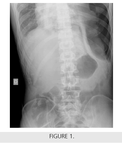
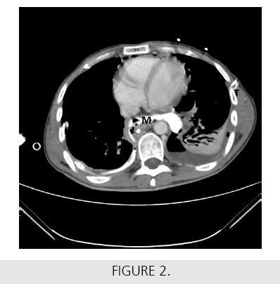
45 year old man, with children cerebral palsy and reflux esophagitis was attended in other hospital due to abdominal pain and fever. Studies done were normal. Two days after, broad spectrum antibiotics were prescribed. Persistent clinical symptoms led to take a plain x-ray of the thoracoabdominal cross, informing a tension pneumothorax with left diaphragm inversion (FIGURE 1). A thoracic tube was inserted and a thoracoabdominal CT was performed afterwards, showing mediastinitis (M) caused by esophageal perforation (FIGURE 2), pneumothorax treated with the thoracic tube (T) and mild pneumoperitoneum. Therefore, the patient was admitted in our hospital and emergency surgery was performed through cervical and abdominal access, undergoing mediastinal drainage, drainage gastrostomy tube, feeding yeyunostomy and cervical monopolar exclusion of the esophagus with stapler. Patient was discharged two months after the operation with normal oral intake. Our case was unique in that a tension pneumothorax was due to esophageal perforation.


