Case Report - Clinical Practice (2022) Volume 19, Issue 1
Successful percutaneous insertion of impella device using axillary artery complicated by radial artery occlusion
- Corresponding Author:
- Jack Xu
Division of Cardiovascular Medicine, University of Arkansas for Medical Sciences, United State
E-mail: jaxu54@gmail.com
Received: 15 December, 2021, Manuscript No. M-49916; Editor assigned: 17 December, 2021, PreQC No. P-49916; Reviewed: 20 December, 2021, QC No. Q-49916; Revised: 02 January, 2022, Manuscript No. R-49916 Published: 10 January, 2022; DOI. 10.37532/ fmcp.2022.19(1).1815-1818
Abstract
A 77-year-old female with a history of Peripheral Vascular Disease (PVD), Aorto Femoral Bypass (AFB), and left subclavian artery stent presented for an elective hip replacement. Her postoperative course was complicated by chest pain with inferolateral ST depressions on ECG. She was found on coronary angiogram to have an 80% calcified lesion in the proximal Left Main Coronary Artery (LM). The decision was made for percutaneous revascularization with the percutaneous placement of an Impella CP via the right axillary artery for mechanical support. Under conscious sedation, the right axillary and radial arteries were accessed under ultrasound guidance. After the Impella sheath was advanced, there was the loss of arterial waveform on the 4 Fr radial sheath. The side port of the Impella sheath was connected to the side port of the radial sheath via an extension tubing, thereby maintaining perfusion of the right hand. The percutaneous coronary intervention was performed on the LM successfully with the Impella being able to be weaned off at the end of the procedure. The present case report highlights the percutaneous utilization of the right axillary artery for mechanical circulatory support in a patient with extensive PVD as well as acute limb ischemia.
Keywords
complex percutaneous coronary intervention, mechanical circulatory support, coronary artery disease, peripheral vascular disease, acute coronary syndrome
Introduction
There has been increasing use of Mechanical Circulatory Support (MCS) devices to guide complex percutaneous coronary intervention or as a bridge to cardiac recovery [1]. Traditionally, the Impella CP is placed via femoral access but in patients with severe peripheral vascular disease, access may be limited [2,3]. If the device is placed in the upper extremity vessels, it usually necessitates a surgical cut-down with a surgeon, anesthesiologist, and a hybrid operating room with fluoroscopic capabilities [4,5]. However, a percutaneous approach can bypass some of those limitations.
Case Presentation
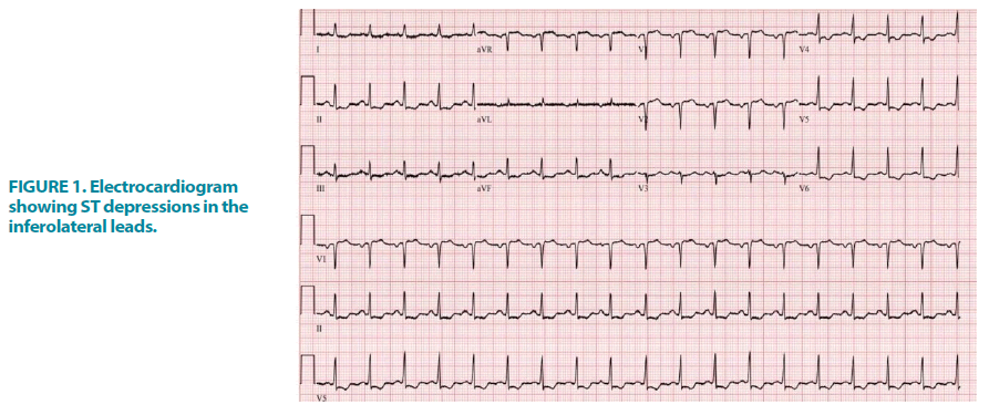
We present a 77-year-old female with a history of non-obstructive coronary artery disease, osteoarthritis, carotid stenosis, Peripheral Vascular Disease (PVD) with an Aorto Femoral Bypass (AFB), and a left subclavian stent who came to the hospital for an elective left hip replacement. She underwent a left total hip replacement but her postoperative course was complicated by chest pain with >1 mm down sloping inferolateral ST depressions on electrocardiogram (FIGURE 1). The patient was deemed to have a non ST-segment elevation myocardial infarction and was scheduled for a left heart catheterization. Before cardiac catheterization, the patient felt numbness in her right foot with nonpalpable pulses in her right lower extremity. Vascular surgery evaluated the patient and was concerned for right limb aortofemoral bypass occlusion. After a multidisciplinary team discussion, the decision was made for a coronary angiogram before vascular revascularization.
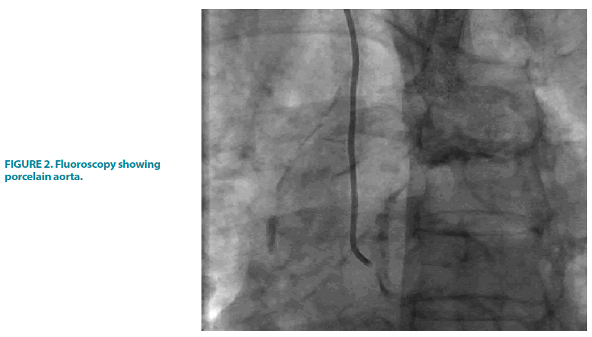
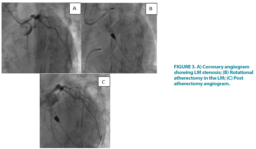
Fluoroscopy of the aorta showed a porcelain aorta (FIGURE 2). The left heart catheterization showed an 80% calcified lesion in the proximal Left Main Coronary Artery (LM) with no other obstructive coronary lesions (FIGURE 3A) and a left ventricular end-diastolic pressure of 31 mm Hg. An echocardiogram showed an ejection fraction of 55%-60% with normal diastolic function, no significant aortic stenosis, and no wall motion abnormalities.
Figure 3: A) Coronary angiogram showing LM stenosis; (B) Rotational atherectomy in the LM; (C) Post atherectomy angiogram.
After the patient was deemed to be at prohibitive surgical risk, the decision was made for percutaneous revascularization with the percutaneous placement of an Impella CP via the right axillary artery for mechanical support. The femoral arteries were not accessed due to the concern of loss of flow in the left AFB in the context of acute thrombotic occlusion of the right AFB. Under conscious sedation, the right axillary and radial arteries were accessed under ultrasound guidance, with preclosure of the puncture site with Perclose ProGlide devices. After the Impella sheath was advanced, there was the loss of arterial waveform on the 4 Fr radial sheath. The side port of the Impella sheath was connected to the side port of the radial sheath via an extension tubing, thereby maintaining perfusion of the right hand. Thereafter, single access was used to insert a 7 Fr sheath into the Impella sheath. Rotational atherectomy was performed using a 1.5 burr, followed by IVUS guided stent placement in the LM, using 4.0 mm × 15 mm drug-eluting stent, which was post dilated with a 4.5 mm × 8 mm non-compliant balloon (FIGURE 3B and FIGURE 3C). Impella was weaned off at the end of the procedure and the Perclose sutures were successfully deployed.
The patient then underwent right AFB limb thrombectomy with systemic heparinization but during the procedure, the patient was hypotensive requiring multiple units of packed red blood cells so an angiogram was done to evaluate for bleeding in the right axillary artery. An angiogram showed extravasation so a covered stent was placed.
Discussion
Percutaneous large-bore access for MCS devices has become a complex and evolving field [1,5]. Patients who need this support oftentimes have multiple comorbidities such as severe PVD. Femoral access is the most common access for Impella devices but in some patients with significant lower extremity PVD, largebore arterial access is not always feasible in the femoral artery [1]. In addition, femoral access necessitates bed rest in patients who could potentially benefit from in-hospital physical therapy.
Impella placement in the upper extremity is usually placed via a surgical cutdown of the axillary artery along with a conduit graft [1,2]. This procedure requires an anesthesiologist, surgeon, and usually a hybrid operating room with fluoroscopic imaging availability [1]. However, a percutaneous approach could enable placement in a standard catheterization laboratory and possibly avoid the need for surgical cut-down or anesthesia [1,4]. It would also allow for patients to participate in rehabilitation.
The present case report highlights the percutaneous utilization of the right axillary artery for MCS in a patient with extensive PVD as well as acute limb ischemia. It also highlights the importance of a multidisciplinary approach when dealing with complex presentations as well as highly morbid patients.
Conclusion
Typically, Impella devices are inserted via femoral access but when severe PVD precludes the use of lower extremity access, we believe that an upper extremity percutaneous approach can be used in a traditional cardiac catheterization laboratory. We believe that ipsilateral radial access is necessary if the large bore sheath occludes flow to the rest of the upper extremity. In our case, we were able to maintain perfusion of the hand using an extension between the Impella sheath and a radial sheath.
References
- McCabe JM, Kaki AA, Pinto DS, et al. Percutaneous axillary access for placement of microaxial ventricular support devices: The Axillary Access Registry to Monitor Safety (ARMS). Circulation Cardio Interven. 14, (2020).
- Lotun K, Shetty R, Patel M, et al. Percutaneous left axillary artery approach for Impella 2.5 liter circulatory support for patients with severe aortoiliac arterial disease undergoing high‐risk percutaneous coronary intervention. J Interv Cardiol. 25, 210-213 (2012).
- Tayal R, Barvalia M, Rana Z, et al. Totally percutaneous insertion and removal of Impella device using axillary artery in the setting of advanced peripheral artery disease. J Invasive Cardiol. 28, 374-380 (2016).
- Mathur M, Hira RS, Smith BM, et al. Fully percutaneous technique for transaxillary implantation of the Impella CP. JACC Cardiovas Interv. 9, 1196-1198 (2016).
- Truong HTD, Hunter G, Lotun K, et al. Insertion of the Impella via the axillary artery for high-risk percutaneous coronary intervention. Cardiovas Revascular Med. 19, 540-544 (2018).


