Clinical images - Imaging in Medicine (2018) Volume 10, Issue 5
Colonic intussusception
Sindhu Kumar*Department of Radiology, University of Virginia Medical Center, Virginia 22901, USA
- Corresponding Author:
- Sindhu Kumar
Department of Radiology
University of Virginia Medical Center
Virginia 22901, USA
E-mail: sindhucumar@gmail.com
Abstract
https://aermech.com https://world-oceans.org https://lline.net https://apecu.org https://febayder.com https://johnbirch.org
Keywords
computed tomography, left lower quadrant
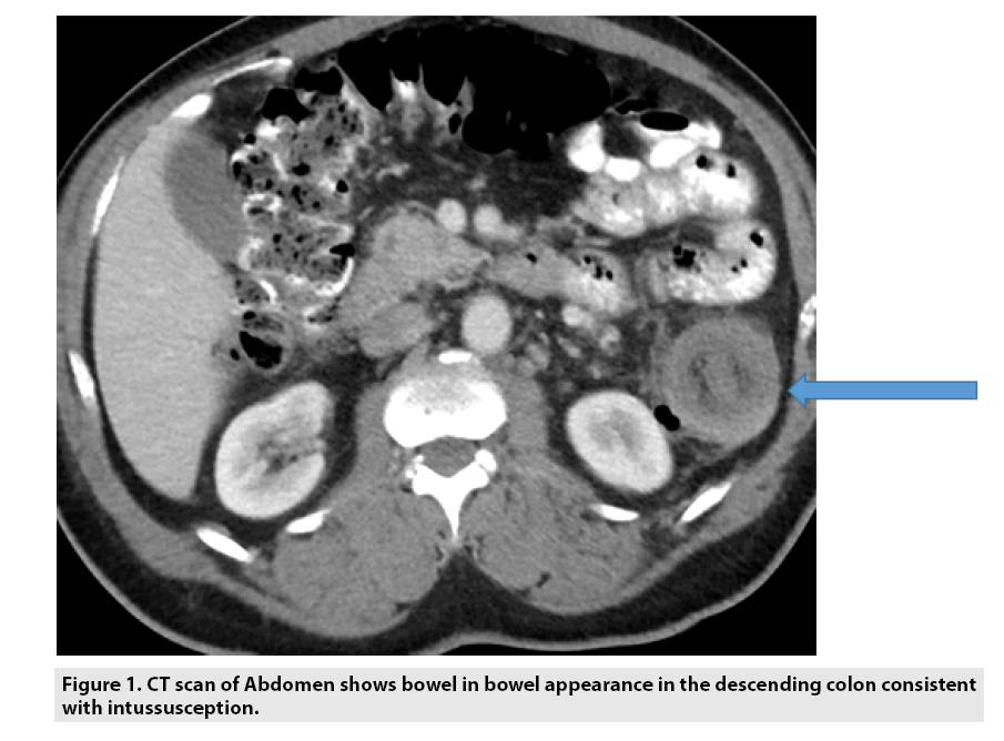
Intussusception is the invagination of a bowel loop with its mesenteric fold (intussusceptum) into the lumen of a contiguous portion of bowel (intussuscipiens) (FIGURE 1). It most frequently involves the small intestine, often incidentally seen on routine CT scans and can be transient. Most common cause are due to neoplasm such as colon cancer, lymphoma or metastases. Most common benign cause is Lipoma.
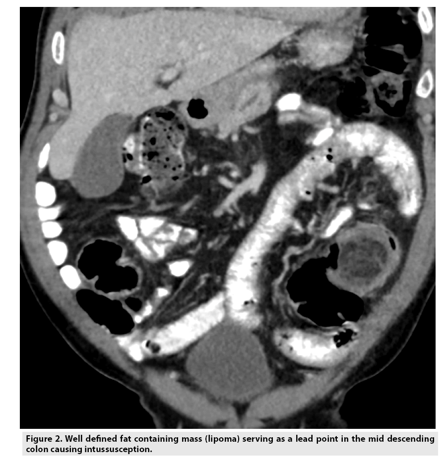
65 y old male presents with LLQ pain and constipation. Colonoscopy was attempted but failed as the scope was not able to traverse the area of concern in the descending colon. Surgery revealed colonic intussusception due to lipoma as the lead point (FIGURE 2).


