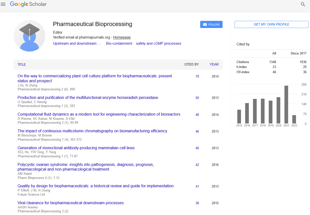
Editorial - Pharmaceutical Bioprocessing (2021) Volume 9, Issue 5
Role of cGMP Regulation in Myocardial Phosphodiesterases
- *Corresponding Author:
- Rene H Wijffels
Department of Food and Bioprocess Engineering Group, Wageningen University and Research Center, Wageningen, Netherlands
E-mail: rene.hwijffels@algemeen.pk.wau.nl
Abstract
PDE1: From cGMP to cAMP modulation within the heart
PDE1 is Ca2+/calmodulin (CaM)-activated and a dual cAMP/cGMP esterase expressed together of three different isoforms. Of these, PDE1A and 1C are expressed within the heart. PDE1A features a 25-fold lower Km for cGMP than Camp, so is more cGMP-selective, whereas PDE1C has an equal affinity for both cyclic nucleotides. PDE1A represents the dominant cardiac isoform in mice and rats, whereas PDE1C predominates in larger mammals like humans, dogs, and rabbits. The N-terminus contains two Ca2+/CaM binding sites and a phosphorylation site at Ser120, the latter modified by PKA in PDE1A and CaMKII in PDE1B to suppress sensitivity to Ca2+/CaM and thus PDE activation. PDE1A and PDE1C expression are up-regulated in hypertrophied rodent hearts and myocardium from human coronary failure. In mice, β-AR stimulated hypertrophy is suppressed by PDE1 inhibition by a mechanism most compatible with cGK1 activation. PDE1A is additionally up-regulated in rodent myo-fibroblasts after myocardial infarct, and PDE1 inhibition blocks expression of pro-fibrotic genes. The anti-fibrotic response involves both cGMP and cAMP signaling.
These data have shifted the main target of PDE1 regulation within the intact heart from one emphasizing cGMP to cAMP signaling, with the latter being coupled to adenosine and not β-AR receptors. The protective effects of A2 receptor stimulation against ischemic injury are documented, and therefore the new data suggest such protection maybe leveraged by PDE1 inhibition. Furthermore, the positive inotropy and lusitropy may differ from that coupled to β-AR-cAMP signaling, with the potential for a special safety/toxicity profile than observed with PDE3 inhibitors. A Phase Ia-IIb clinical test testing safety and hemodynamic responses to single dose of ITI-214 in patients with dilated coronary failure.
PDE2: Signaling ambiguities and alternative roles
PDE2 may be a dual substrate phosphodiesterase that hydrolyzes cAMP and cGMP at similar maximal rates. One gene Pde2a gives rise to 3 isoforms (PDE2A1, 2A2, and 2A3) which are differentially localized in cytosol, membrane, and mitochondrial sub-domains. A primary characteristic of PDE2A is its allosteric activation by binding of cGMP to GAF domains residing within the N-terminus that augments catalytic activity of cAMP by 10-30 fold. The reverse, cAMP mediated activation for cGMP hydrolysis, doesn’t occur given the low affinity of the GAF domain for cAMP. This positions the PDE for particular relevance to cGMP/cAMP crosstalk.
The role of PDE2 in hydrolyzing cGMP generated by nitric-oxide stimulation of GC-1 has eluded detection by similar fluorescent sensor methods, presumably primarily thanks to insufficient sensitivity to the low amplitude cGMP signal. However, functional data support such interaction. For instance, NO-stimulated cGMP, coupled to activation of the β3-adrenergic receptor, was shown to activate PDE2 (cGMP binding to regulatory GAF domains) in neonatal cardiomyocytes. This enhanced its hydrolysis of cAMP and thereby countered catecholamine stimulated contraction. PDE2 inhibition also enhances cAMP-induced dilation induced by prostaglandin I2, supporting a task for cAMP hydrolysis in plant tissue. yet one more study reported anti-hypertrophic effects from PDE2 inhibition in isolated neonatal cardiomyocytes exposed to angiotensin II , but this was observed when cGMP was co-stimulated using an NO-donor but not by a NPR1 agonist (ANP or BNP). Similarly, up-regulation of PDE2 (genetic overexpression) in fibroblasts didn’t alter cGMP stimulated by NP agonism, but had a small significant effect upon NO stimulation. These latter studies are hard to reconcile with earlier data coupling PDE2 to NPR1 signaling, but they’ll reveal a critical role of experimental conditions and cAMP/cGMP balance.
PDE3 – Isoform-specific signaling for therapeutic targeting
PDE3 is that the other dual-substrate phosphodiesterase proposed to manage cGMP within the heart. While its affinity for cAMP and cGMP is analogous, it’s a better employee turnover for cAMP, providing its primary regulatory footprint in larger mammalian hearts including human. By competitive binding at the catalytic site, cAMP hydrolysis is inhibited by cGMP, yielding a mechanism for cGMP-dependent contractility augmentation at low levels of cGMP44. The PDE3 enzymes are transcribed from two genes, PDE3A and PDE3B. PDE3A exists as three isoforms that change by alternative transcription and translation start sites, while PDE3B exists as just one isoform. All of the isoforms differ in their N-terminus where there are hydrophobic loops (PDE3A1 and PDE3B) to mediate lipid membrane insertion, and phosphorylation sites that promote protein-protein interactions. The latter is vital for determining the intracellular localization of the isoforms. Especially, PDE3A has been found to complex with PI3Kγ and SERCA2A, and is especially localized within the cardiomyocyte at the sarcoplasmic reticulum. Against this, PDE3B localizes to myocyte T-tubule membranes.
PDE5 – Myocardial regulation and compartmentation
PDE5A may be a cGMP-specific PDE expressed together of three isoforms PDE5A1, A2, and A3, all three present in human and mouse and ranging in their N-terminus. In vitro biochemical function is analogous among the isoforms, though there are organ-specific differences in expression, and evidence that this might also influence subcellular localization66. PDE5A cGMP-catalytic activity is stimulated by cGMP binding to GAF regulatory domains and by phosphorylation by PKG at serine S102 in human (S92 in mouse). In cardiac myocytes, localization of PDE5A appears during a striated banding pattern that co-localizes with the z-disk protein α-actinin. This localization normally favors hydrolysis of cGMP generated by the nitric oxide-GC-1α pathway as compared to natriuretic peptide (GC-A or GC-B) pathway. However, studies in both adult canine and murine cardiomyocytes found this localization changes to a more diffuse cytosolic one in models of coronary failure and hypertrophy, or if NOS3 is pharmacologically or genetically suppressed. Interestingly, its normal z-disk localization is restored even in hearts with chronic NOS inhibition by directly co-activating GC1α to get cGMP. The precise mechanism for altered intracellular PDE5A localization with heart condition remains unknown.
PDE9A - Differential Effector of NO versus NP-generated Cgmp
PDE9A may be a cGMP-specific PDE encoded by one gene that’s then alternatively spliced to supply multiple isoforms. It’s expressed throughout the body, including lung, kidney, heart, and striated muscle, but most prominently in cerebellar Purkinje neurons and at lower levels in cortex, hippocampus, and striatum. Brain expression is assumed to play a task in synaptic plasticity, and studies so far have focused on this role and potential therapeutic use for disorders with cognitive disease like Alzheimer’s and schizophrenia. There’s isoform specific expression in several tissues, with brain expressing PDE9A6/13 and three higher relative molecular mass isoforms – PDE9x-100, −120, and −175. These aren’t found within the heart, which instead expresses PDE9A2, and 9A9. Isoform-specific functional differences have yet to be identified, and inhibitors of the enzyme don’t differentiate between them.