Case Report - International Journal of Clinical Rheumatology (2024) Volume 19, Issue 5
Maffucci syndrome: a rare and unknown
Mxxk Djennane1,2*, Rima Hassani2, Mbriam Amal Ifticene2 and Linda igueni1,2
1Faculty of Medicine, Centre hospitalier universitaire, Algeria
2Rheumatology departments, Centre hospitalier universitaireTizi Ouzou, Algeria
- *Corresponding Author:
- Mxxk Djennane
Faculty of Medicine, Centre hospitalier universitaire, Algeria and Rheumatology departments, Centre hospitalier universitaireTizi Ouzou, Algeria
E-mail: Malik.djennane@hotmail.com
Received: 16-Mar-2024, Manuscript No. fmijcr-24-129861; Editor assigned: 18- Mar-2024, Pre-QC No. fmijcr-24-129861 (PQ); Reviewed: 01-Apr-2024, QC No. fmijcr-24-129861; Revised: 01-May-2024, Manuscript No. fmijcr-24-129861 (R); Published: 11-May-2024, DOI: 10.37532/1758-4272.2024.19(5).56-59
Abstract
Maffuci’s syndrome is a rare non hereditary mesodermal dysplasia, classically defined as the association of multiples enchondromas and soft tissue hemangiomas. A systemic regular clinical and radiological evaluation should be considered because this syndrome is related to a high incidence if malignant transformation.
Keywords
Maffuci’s syndrome • Hemangiomas • Enchondrom • Sarcomatous transformation
Introduction
Angiochondromatosis, or Maffucci syndrome (SM), is a congenital, non-hereditary condition first described by Maffucci in 1881 [1]. It is classically defined by the association of multiple soft tissue hemangiomas and chondromas predominantly located in the phalanges [2]. It is a rare condition, approximately 200 cases have been reported in the world literature [3,4]. The risk of association with malignant or benign tumors is considerable [5].
We report the observation of one case.
Observation
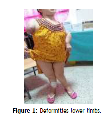
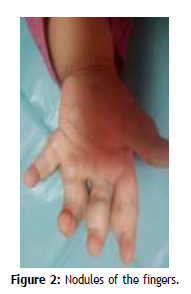
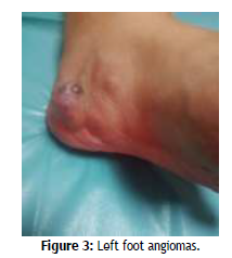
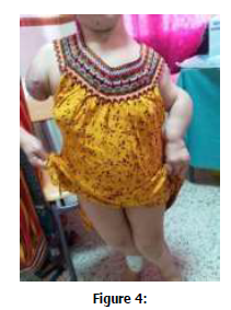
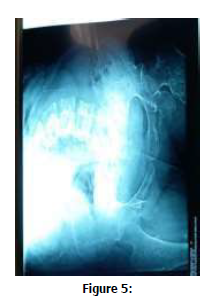
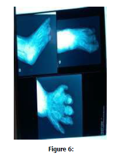
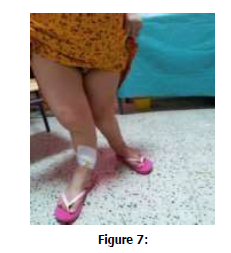
B.K, aged 29, from a consanguineous marriage, has progressively worsening bone deformities that have progressed since the age of 2 and gradually increase in number and size. In the history, there is an amputation of the upper right limb 6 months ago, the cause of which has not been documented. No similar cases in the family. The osteoarticular examination reveals severe dorsolumbar scoliosis, multiple painless rounded formations predominantly on the fingers (Figure 1), toes and deforming the left forefoot (Figure 2) as well as deformities in the long bones as well as a marked shortening of the upper and lower limbs associated with 3 angiomas in the left foot (Figure 3). In biology, there is no inflammatory syndrome and the rest of the workup is without abnormality. The standard x-ray confirmed the severe scoliosis and demonstrated poorly limited lacunar lesions blowing the cortex on the hands (Figure 4), femur, tibia, fibula (Figure 5), and feet (Figure 6). As well as lumbar scoliosis and pelvic deformity (Figure 7). Ultrasound of the soft tissues at the level of the swelling: slightly heterogeneous hypoechoic tissue nodules, vascularized with well-limited color Doppler, measuring for the largest 20/12 mm. The remainder of the physical examination for bone, neurological or ocular malformations was unremarkable.
Discussion
Maffucci syndrome is a very rare dysplastic disorder of the mesoderm. About 250 cases have been reported in the literature so far. MS affects both men and women, without ethnic or geographic predilection [3]. This condition is characterized by the association of benign cartilage tumors similar to the enchondromas of Ollier's disease, which are predominantly located in the phalanges and long bones, and cutaneous hemangiomatosis [6]. It results from mesodermal dysembryoplasia, which explains its dual vascular and cartilaginous component. Vascular lesions can be of three types: cavernous hemangiomas, phlebectasias or lymphangiomas [1]. The multiple enchondromas of Maffucci syndrome are the cause of benign enlargements of the cartilage, most often present in the phalanges and long bones but which can appear anywhere. They can manifest as painless swelling of the fingers or feet or a pathological fracture and lead to significant deformities with a predilection for the phalanges, metacarpus, metatarsus and long bones of the legs and forearms [4] .Capillary malformations are usually manifested by the presence of irregularly shaped, protruding and dark blue subcutaneous nodules at the distal ends but they can appear anywhere. There may be venous and lymphatic malformations may appear. Skeletal and vascular lesions are usually asymmetric and may be progressive. About 30-40% of enchondromas evolve into chondrosarcomas. The syndrome can be associated with other benign or malignant tumors (goiter, parathyroid adenoma, pituitary adenoma, adrenal tumor, ovarian tumor, breast cancer, or astrocytoma). An important question concerns the follow-up of patients with Maffucci syndrome, namely the early detection of malignant tumors. In order to identify chondrosarcomas, Vedegaal and his colleagues proposed technetium scans in patients with more than one enchondroma. Radiographs of each enchondroma have been recommended to provide a basis for future comparison [7]. To identify non-skeletal neoplasms, some authors have recommended a brain or abdominal CT scan when neurological or abdominal symptoms appear [7]. The diagnosis is based on the presence of clinical and radiological manifestations. Management consists of relieving symptoms and early detection of malignant tumors. No treatment is recommended for asymptomatic patients. Regular examinations by an orthopedic surgeon and a dermatologist in order to assess the evolution of bone and skin lesions are necessary.
Conflicts of Interest
The authors declare no conflicts of interest.
References
- Lee NH, Choi EH, Choi WK et al. Maffucci’s syndrome with oral and intestinal haemangioma. Br J Dermatol 140, 968-9 (1999).
- Faik A, Allali F, El Hassani S et al. Maffucci’s syndrome: A case report. Clin Rheumatol 25, 88-91 (2006).
- Sang Mi Lee, Jong Jin Lee, Yu Kyeong et al. Bone Scintigraphy Findings of A Case with Maffucci’s Syndrome. Nucl Med Mol Imaging 44, 150–153 (2010).
- Nutan F. Maffucci syndrome in a 10-year old boy. Contemporary Pediatrics 51-54 (2011).
- Yazidi A, Benzekri L, Senouci K et al. Syndrome de Maffuci avec carcinome épidermoïde du cavum. Ann Dermatol Venereol 125, 50-51 (1998).
- Lissa FC, Argente JS, Antunes GN et al. Maffucci syndrome and soft tissue sarcoma: a case report. Int Semin Surg Oncol 13, 6-2 (2009).
- Verdegaal SH, Bovee JV, Pansuriya TC et al. Incidence, predictive factors, and prognosis of chondrosarcoma in patients with Ollier disease and Ma ucci syndrome: an international multicenter study of 161 patients. Oncologist. 16, 1771–1779 (2011).
- Benbouazza K, El Hassani S, Hassikou H et al. Multiple enchondromatosis. Joint Bone Spine 69, 236-239 (2002).
Indexed at, Google Scholar, Crossref
Indexed at, Google Scholar, Crossref
Indexed at, Google Scholar, Crossref
Indexed at, Google Scholar, Crossref
Indexed at, Google Scholar, Crossref
