Case Series - Interventional Cardiology (2023)
Coronary stent loss-outcomes and their management
- Corresponding Author:
- Chetana Krishnegowda
Department of cardiology, Sri Jayadeva Institute of Cardiovascular Sciences and Research, Karnataka, India,
E-mail: chetana88@gmail.com
Received date: 18-May-2023, Manuscript No. FMIC-23-99209; Editor assigned: 22-May-2023, PreQC No. FMIC-23-99209 (PQ); Reviewed date: 06-Jun-2023, QC No. FMIC-23-99209; Revised date: 13-Jun-2023, Manuscript No. FMIC-23-99209 (R); Published date: 21-Jun-2023, DOI: 10.37532/1755- 5310.2023.15(S17).424
Abstract
Coronary stent loss and embolization is an infrequent complication in the current era of percutaneous coronary interventions. The incidence of stent loss has significantly reduced owing to the factory crimping of the newer generation stents. It is often benign and managed conservatively by retrieval, deployment or crushing of the stent, rarely requiring surgical intervention. However, lethal complications can occur if the stent embolizes into the vital organs.
Keywords
Coronary stent loss • Stent dislodgement • Stent embolization
Introduction
Coronary stents are the most commonly embolized devices, with an incidence of approximately 0.9%-8.3%, during Percutaneous Coronary Interventions (PCI).Stent dislodgement was more frequent in the past when stents were manually crimped onto the balloon [1]. Current generation of balloon-mounted stents with better radio-opacity have reduced the incidence of stent dislodgement. Although rare, stent dislodgement can be hazardous in some circumstances, where it may result in cerebral embolism or intracoronary thrombosis and lead to fatal complications like Cerebrovascular Accident (CVA) and Myocardial Infarction (MI). Several techniques of stent retrieval or exclusion have been described in the past. The simplest and the most common practiced method of stent retrieval is using a snaring device [2-6]. We report 4 cases of Coronary Stent Loss (CSL) managed successfully with snaring technique.
Case Presentation
Case 1
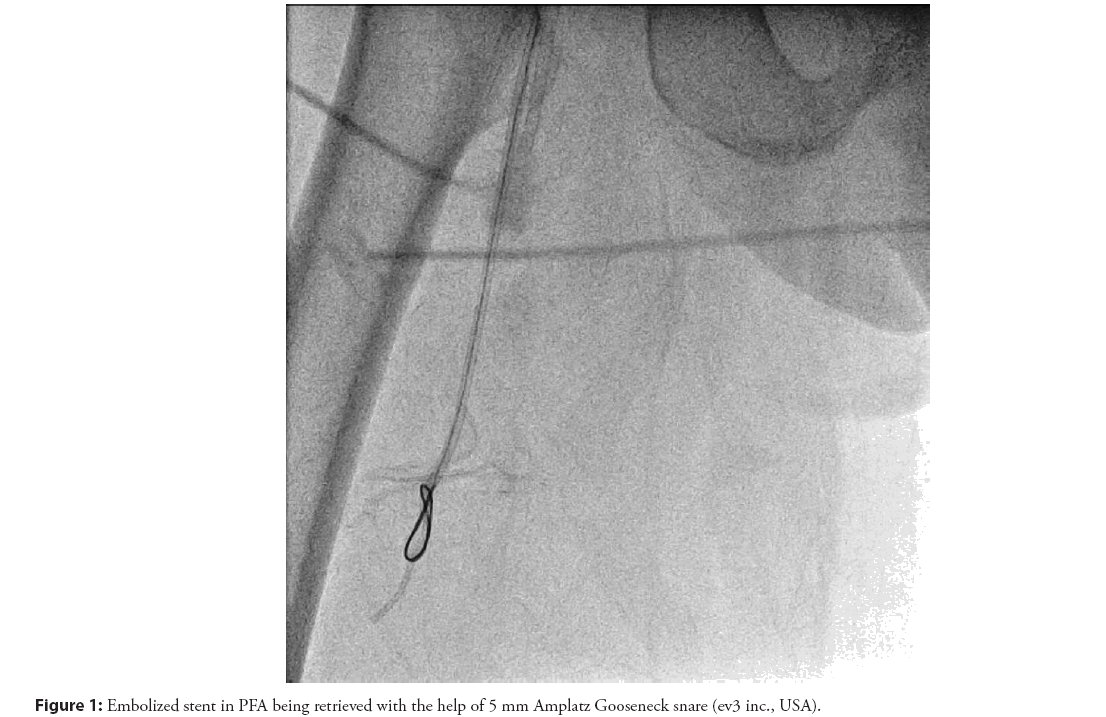
A 72 year old female presented with chest pain of 4 days duration. Electrocardiogram (ECG) showed q waves in the anterior leads. Echocardiography showed wall motion abnormalities in the Left Anterior Descending (LAD) territory with good Left Ventricular (LV) function (ejection fraction 55%). Due to ongoing angina, she was subjected for Coronary Angiogram (CAG) which showed 90% stenosis in mid segment of LAD. Left Circumflex (LCX) and Right Coronary Artery (RCA) were normal. PCI of LAD was planned. Right femoral arterial access was taken. The left main ostium was cannulated with 6 F Extra back up (EBU 3.5, Medtronic, Minneapolis, USA) catheter. LAD was wired with 0.014” Cougar XT guidewire and predilated with 2.5 × 12 mm semi compliant balloon @12 atmospheres (atm). 2.75 × 28 mm Sirolimus Drug Eluting Stent (DES) was taken. The stent got stuck at the proximal LAD lesion and was unable to cross the target lesion. Hence predilation with a larger balloon for lesion modification was planned. During attempts to withdrawal of the stuck stent, stent got partially dislodged from the underlying balloon. Since part of the stent balloon was still inside the stent, with partial inflation of the balloon the whole catheterization system was pulled en bloc into the abdominal aorta. While removing the system through the femoral artery sheath the stent embolized into the Profunda Femoris Artery (PFA). Through the contralateral femoral artery access, the embolized stent in the PFA was reached with 5 F Multipurpose (MP) catheter (Cordis Corporation, USA)-Terumo wire combination and then with the help of 5 mm Amplatz Gooseneck snare (ev3 inc., USA) stent was successfully retrieved (Figure 1).
Case 2
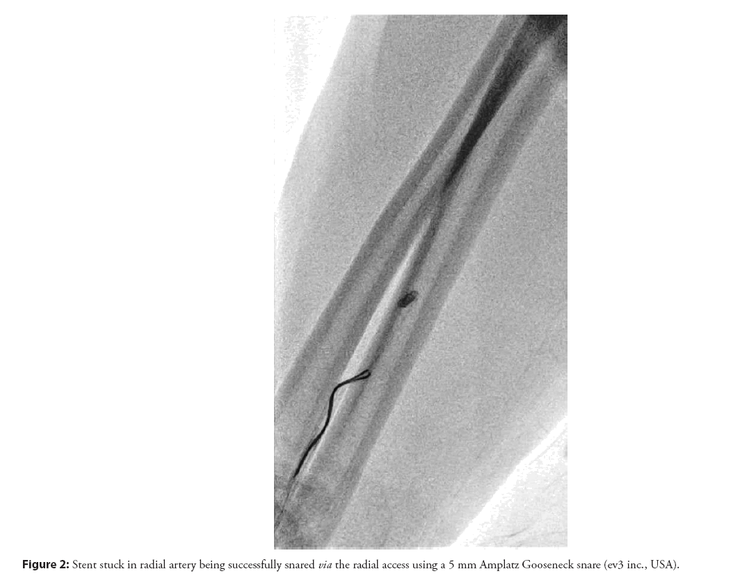
A 72 year old female with history of type 2 diabetes mellitus and hypertension presented with inferior wall MI. Patient was taken for CAG. RCA was totally occluded in the proximal segment. A hydrophilic wire (Hi-Torque Whisper MS, Abbott) was used to cross the lesion. Predilatation was done with 2.5 × 12 mm Sprinter® balloon (Medtronic Inc.) at 10 atm. A 3 × 40 mm Sirolimus DES was taken. As the stent failed to track across the diseased segments, stent withdrawal from the vessel was attempted. But the stent got dislodged from the underlying balloon partially. The whole system was retrieved out into the right subclavian artery and further into the right brachial artery. As the system was retrieved through the radial sheath, the stent got completely dislodged from the system and was freely floating in the radial artery. The stent was successfully snared via the radial access using a 5 mm Amplatz Gooseneck snare (ev3 inc., USA) through the 5F MP catheter (Figure 2). PCI of RCA was then completed through the same radial artery access successfully.
Case 3
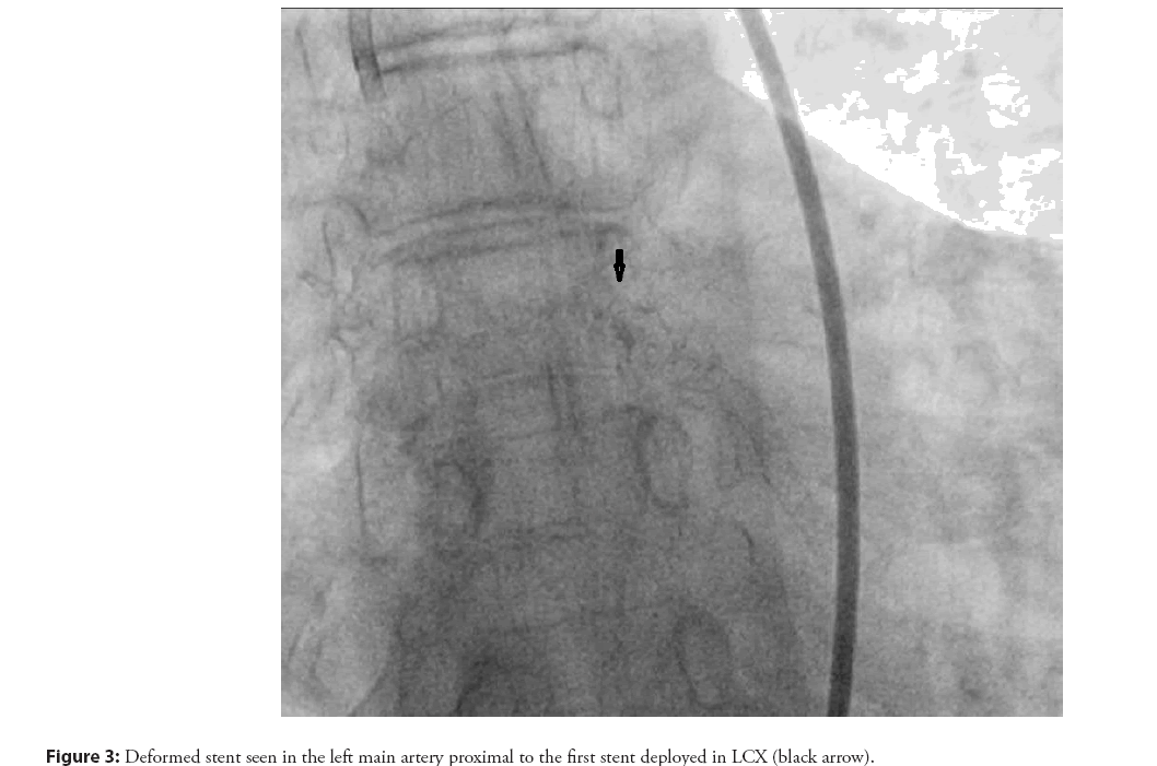
A 62 year old female, presented with Non ST Elevation Myocardial Infarction (NSTEMI). CAG showed single vessel disease. LCX was dominant and had a subtotal occlusion. PCI was planned for LCX. The lesion was crossed with a 0.014 inch Balance Middle Weight (BMW) coronary wire. Adequate predilatation was done with 2.5 × 12 mm semi-compliant balloon at 14 atm. A 2.75 × 32 mm Sirolimus DES was deployed at 12 atm and post dilatation was done with 3 × 12 mm 1:1 non-compliant balloon (Medtronic, Minneapolis, USA) at 18 atm. Check shoot following post dilatation showed edge dissection and distal TIMI 1 flow. So covering the dissected segment with a 2.5 × 18 mm Sirolimus DES was planned. But the second stent could not be negotiated through the already deployed stent in the proximal LCX even after repeated attempts. So as the undeployed stent was withdrawn into the catheter, it got dislodged from the balloon partially. Stent balloon was inflated at 4 atm and the whole system was removed en bloc. But a stent tine had got caught in the struts of the previously deployed stent in proximal LCX and hence when the whole system was withdrawn, the undeployed stent got stretched and proximal end of it was found to be floating out of the left main ostium into the aortic cusp (Figure 3). Subsequently, keeping the guide catheter (EBU 3.5, Medtronic, and Minneapolis, USA) close to the left main ostium, and using a Gooseneck snare (ES-10, AndraTec), the deformed stent was successfully retrieved in toto.
Case 4
A 70 year old male patient, hypertensive and diabetic, presented with NSTEMI. His angiogram revealed a severe calcified stenosis of 90% in the mid-RCA segment. The RCA ostium was engaged with 6F Judkins Right guide catheter and vessel was prepared for stent deployment. A 3 × 32 mm Sirolimus DES was taken into the RCA. The stent could not be negotiated across the lesion easily. During attempts to negotiate the stent to the desired segment the stent got dislodged partially from the balloon. Fortunately, the guidewire was in situ, so we took a guideliner as close to the stent as possible and with the help of FAndraTec Exeter micro retreiver removed the system en bloc [4]. Later, the vessel was rewired and two smaller stents were taken and successfully deployed across the lesion.
Results and Discussion
CSL complications, although rare (<1%), have not been completely eliminated. Stent dislodgement can be explained by various mechanisms in different scenarios. Stent entrapment is one of the mechanisms where dissociation from the stent balloon occurs while pulling the stent into the guiding catheter. Stent push- back is another mechanism where dissociation occurs during the insertion of the stent through the lesion. In our series the first three cases involved stent entrapment. Fourth case involved stent push back as the mechanism. Factors which increase the chances of stent detachment from the balloon catheter within a coronary artery as described in previous case reports include tortuosity, calcification, and passage through a previous stent [1]. In our case series, all four cases involved tortuous vessels and severely calcified lesions, which were the major contributing factors for stent dislodgement.
Various management options can be applied in different scenarios. A number of stent retrieval techniques have been described in the past. These include two twisted guide wires or braiding technique, use of loop snares, small-balloon technique and lastly, the stent- crush exclusion technique, whereby a second stent is used to crush the detached stent along the wall of the coronary artery [6-9].
In case 1, though the stent was successfully retrieved the other treatment option would have been to leave the stent in peripheral vessel as it was a small artery. Similarly in case 2, conservative approach of leaving the stent behind in peripheral vessel was a decent option as radial artery occlusion is rarely accompanied by hand ischemia, because of the dual blood supply of the hand and rich network of collateral circulation. Alternatively radial artery cut down could have been done, as the artery is superficial and easily accessible.
Key principles to be followed during coronary stent loss scenarios are:
1) Maintain the guidewire position inside the dislodged stent as much as possible as it helps in retrieval of the stent.
2) Stent should be brought to the distal part of abdominal aorta or the iliac artery bypassing the arch vessels and major branches.
3) Get another access to ease stent retrival especially if the stent is disloged in the lower limb arteries.
4) Gentle retraction back into the guide catheter keeping the guide coaxial to the vessel lumen prevents majority of stent embolization.
5) Sincere attempt to retrieve the embolized stent to be made before resorting to conservative management.
6) Ensure adequate anticoagulation throughout the procedure
Conclusion
No specific treatment fits all case scenarios. Each case of CSL needs to be assessed individually. Position of the guidewire is crucial in the choice of the retrieval technique. Although most cases of stent dislodgement are benign and amenable for retrieval, rarely when stent embolizes to cerebral/carotid arteries, fatal complications like CVA, MI and death can occur.
References
- Brilakis ES, Best PJM, Elesber AA, et al. Incidence, retrieval methods, and outcomes of stent loss during percutaneous coronary intervention. Catheter Cardiovasc Interv. 66(3): 333-340 (2005).
- Rozenman Y, Burstein M, Hasin Y, et al. Retrieval of occluding unexpanded Palmaz-Schatz stent from a saphenous aorto-coronary vein graft. Cathet Cardiovasc Diagn. 34(2): 159-161 (1995).
- Veldhuijzen FL, Bonnier HJ, Michels HR, et al. Retrieval of undeployed stents from the right coronary artery: Report of two cases. Cathet Cardiovasc Diagn, 30(3): 245-248 (1993).
- Elsner M, Peifer A, Kasper W, et al. Intracoronary loss of balloon-mounted stents: Successful retrieval with a 2 mm-microsnare-device. Cathet Cardiovasc Diagn. 39(3): 271-276 (1996).
- Foster-Smith KW, Garratt KN, Higano ST et al. Retrieval techniques for managing flexible intracoronary stent misplacement. Cathet Cardiovasc Diagn. 30(1): 63-68 (1993).
- Wongpraparut N, Yalamachili V, Leesar MA, et al. Novel implication of combined stent crushing and intravascular ultrasound for dislodged stents. J Invasive Cardiol. 16(8): 445-446 (2004).
- Alomar ME, Michael TT, Patel VG, et al. Stent loss and retrieval during percutaneous coronary interventions: A systematic review and meta analysis. J Invasive Cardiol. 25(12): 637-641 (2013).
- Colkesen AY, Baltali M, Acil T, et al. Coronary and systemic stent embolization during percutaneous coronary interventions: A single center experience. Int Heart J. 48(2): 129-136 (2007).
- Bolte J, Neumann U, Pfafferott C, et al. Incidence, management, and outcome of stent loss during intracoronary stenting. Am J Cardiol. 88(5): 565-567 (2001).


