Case Report - Interventional Cardiology (2014) Volume 6, Issue 5
Successful non-surgical management of entrapped hydrophilic guidewire during percutaneous coronary intervention
- Corresponding Author:
- Sanjay C Porwal
KLEs Dr Prabhakar Kore Hospital & Medical Research Centre, Belgaum, Karnataka, India
Tel: +91 944 980 0639
E-mail: drsanjayporwal@gmail.com
Abstract
Guidewire breaking or entrapment during the procedure is a rare but well-reported complication of percutaneous coronary intervention. The entrapped guidewire can be managed through intervention, surgery or conservatively depending upon the clinical situation and position of the guidewire. However, the entrapped guidewire leads to serious complications such as thrombosis, occlusion of the coronary vessel or systemic embolism. As far as the authors’ knowledge is concerned, there is no guideline available for optimal management of entrapped guidewire even though such cases have been reported since the beginning of percutaneous intervention era. Here the authors present a case of hydrophilic guidewire entrapment where a different technique for removal was used.
Guidewire breaking or entrapment during the procedure is a rare but well-reported complication of percutaneous coronary intervention. The entrapped guidewire can be managed through intervention, surgery or conservatively depending upon the clinical situation and position of the guidewire. However, the entrapped guidewire leads to serious complications such as thrombosis, occlusion of the coronary vessel or systemic embolism. As far as the authors’ knowledge is concerned, there is no guideline available for optimal management of entrapped guidewire even though such cases have been reported since the beginning of percutaneous intervention era. Here the authors present a case of hydrophilic guidewire entrapment where a different technique for removal was used.
Background
Percutaneous transluminal coronary angioplasty (PTCA) is an effective and relatively safe procedure for the relief of acute coronary syndrome. One of the very rare complications of PTCA is entrapment of catheter remnants, for example, the guidewire, stent or rotablator in coronary arteries [1]. Cases of fragmentation and entrapment of guidewire have been reported since the beginning of the coronary angioplasty era (in the late 1980s) [2]. Depending upon the clinical situation and the position of the entrapped guidewire, physicians use different approaches, for example, surgical removal, percutaneous extrication of the guidewire or conservative management. Although few physicians preferred to manage guidewire conservatively (leaving the entrapped guidewire within the coronary bed and following the patients with systemic anticoagulants) [3], entrapment of guidewire in coronary vasculature may lead to thromboembolic occlusion and thereby acute ischemic events in some cases [4]. The authors report here case of a patient who experienced this rare complication of PCI – entrapment of hydrophilic guidewire after deployment of drug-eluting stents in the left anterior descending coronary artery (LAD). The authors used a different approach for the removal of the stuck guidewire to avoid post-procedural complications due to remnants of the guidewire.
Case report
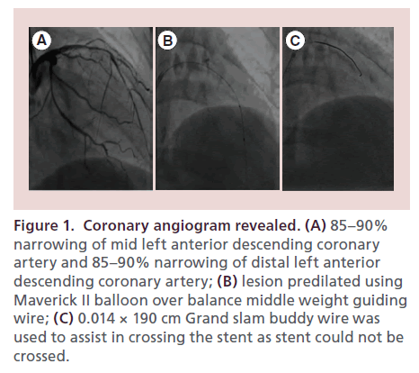
A 41-year-old female attended the clinic with the history of chest pain. She described chest discomfort as being sensation of tightness or squeezing. Hypertension and diabetes, risk factors for developing acute coronary syndrome, were present in the patient. Evaluation of the patient was suggestive of anterior wall myocardial ischemia. Coronary angiography (Figure 1A) revealed double vessel disease, 85–90% narrowing of mid LAD, 85–90% narrowing of distal LAD and 85–90% narrowing of long segment of mid right coronary artery. Coronary angioplasty was planned and it was decided to implant drug-eluting stents in the LAD and right coronary artery.
Figure 1. Coronary angiogram revealed. (A) 85–90% narrowing of mid left anterior descending coronary artery and 85–90% narrowing of distal left anterior descending coronary artery; (B) lesion predilated using Maverick II balloon over balance middle weight guiding wire; (C) 0.014 × 190 cm Grand slam buddy wire was used to assist in crossing the stent as stent could not be crossed.
Left coronary ostium was selectively engaged by using Launcher® 6 F (EBU 3.5) guiding catheter (Medtronic Vascular, MN, USA) through right transfemoral route. We attempted to cross the mid-LAD long segment 85–90% lesion and distal and mid-third 85–90% lesion by using 0.014 × 190 cm hi-torque balance middle weight (BMW) guidewire (Abbott Vascular, USA). As LAD was diffusely narrowed, serial predilatation of the lesion was done using 2.5 × 15 mm Maverick II balloon (Boston Scientific Corp., USA) at 6–10 atmospheric (atm) pressure for 30 s over the BMW guidewire (Figure 1B). Post-balloon check angiography showed 30–40% residual lesion in the LAD. The long stent could not be crossed using BMW guide wire hence 0.014 × 190 cm Grand Slam buddy hydrophilic wire (Abbott Vascular, USA) was used to assist in crossing the stent (Figure 1C).
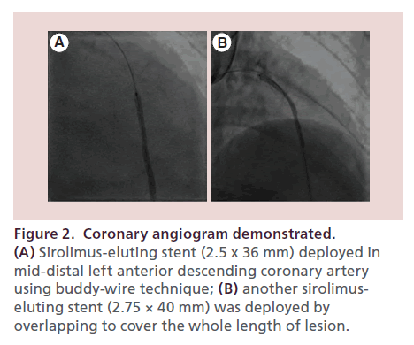
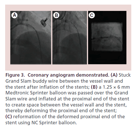
Two sirolimus-eluting overlapping stents were deployed in mid-to-distal LAD and proximal-tomid LAD lesions using balloons (2.5 × 36 mm and 2.75 × 40 mm) at 10–14 atm pressure for 30 s to cover the entire length of the lesion (Figure 2A & B). But unfortunately the removal of the Grand Slam buddy wire was overlooked. Post deployment, it was observed that the buddy wire was stuck between the stent and the vessel wall (Figure 3A). Various attempts were made to remove the buddy wire, such as pulling the wire, use of noninflated small balloon (as support on the buddy wire) to pull the wire while flushing with saline. However, despite all attempts, it was unable to be removed.
Figure 2. Coronary angiogram demonstrated. (A) Sirolimus-eluting stent (2.5 x 36 mm) deployed in mid-distal left anterior descending coronary artery using buddy-wire technique; (B) another sirolimuseluting stent (2.75 × 40 mm) was deployed by overlapping to cover the whole length of lesion.
Finally we tried to create space between the inflated stent struts and the vessel wall to remove entrapped buddy wire using a 1.25 × 6 mm Medtronic sprinter balloon (Medtronic Vascular) (Figure 3B). The guiding catheter was deep-throated to the proximal end of the stent and then the entire system, comprising of the balloon, guide wire and the guiding catheter, was removed.
Figure 3. Coronary angiogram demonstrated. (A) Stuck Grand Slam buddy wire between the vessel wall and the stent after inflation of the stents; (B) a 1.25 × 6 mm Medtronic Sprinter balloon was passed over the Grand Slam wire and inflated at the proximal end of the stent to create space between the vessel wall and the stent, thereby deforming the proximal end of the stent; (C) reformation of the deformed proximal end of the stent using NC Sprinter balloon.
During this process, the proximal end of the deployed stent was deformed. So, the entire stented segment was recrossed with the BMW guiding wire. In-stent dilatation of the deformed segment of the proximal end of the stent was done using NC sprinter balloon (2.75 × 12 mm) at 16–18 atm pressure for 30 s (Figure 3C). Finally the stent regained its reformed structure. The stay of the patient in hospital was uneventful and we did not observe any adverse event related to the stent during 6-month follow-up.
Discussion
One of the rare but serious complications of PTCA is entrapment of catheter remnants in coronary vasculature. The possible complications of entrapped devices include thrombosis, emboli, sepsis, vessel dissection and perforation [5]. Intracoronary thrombosis as a result of entrapped remnant may lead to myocardial ischemia, infarction or arrhythmia and thereby prove life threatening [4,6]. Such a case was reported by Giammarco et al. [4]. They reported a case of 75-year-old patient who underwent PTCA in view of stenosis (60%) in LAD. The patient repeatedly experienced acute myocardial infarction after the procedure. The patient again underwent PTCA and complete occlusion of LAD was found due to a fragment of dilator used in the previous procedure. Various approaches for the retrieval of catheter remnants have been used by cardiologists. Initial case reports demonstrated the role of emergent cardiac surgery for retrieval of catheter remnants. Another alternative is percutaneous retrieval of the remnants using snares, balloons, forceps and bioptomes. There are several techniques described in the literature, that is, twisting two guidewires around the retained guidewire portion followed by removal through the proximal portion of the vessel, inflation of the balloon (at the distal part of the entrapped device) and using it to push the fragment into the proximal portion of the vessel [6,7].
Depending on the position of the catheter remnant and the clinical condition of the patient, the patient is managed conservatively with anticoagulant therapy – leaving the catheter remnant in situ [5]. Although several case reports have reported event-free survival of the patient [8,9], some case reports demonstrated progressive stenosis of the coronary segment that contained catheter remnants [10]. So in this case, the authors tried their best to remove the catheter remnant and avoid long-term complications.
In this case report, the authors present a case of a patient who underwent PTCA in view of double vessel disease (stenosis in mid right coronary artery and distal and mid LAD) after written informed consent of the patient. There was 30–40% stenosis observed in post-balloon check angiography. As hydrophilic guidewire performed excellently in crossing tight and complex lesions [11], hydrophilic wire was used to assist in crossing a long stent. A drug-eluting stent was deployed to cover the entire lesion. But the authors overlooked removal of the hydrophilic guidewire which was used to cross the torturous lesion. The wire stuck between the deployed stent and vessel wall.
To avoid the complications by keeping the guidewire inside the coronary vasculature, various attempts were tried. Usually, the buddy wire can easily be removed even after inflation of the stent. However, in this case the segment was about 60–70 mm in length and the hydrophilic nature of the buddy wire lead to the entrapment. The guidewire remained stuck even after several attempts. Very-low-profile balloon was used. Non-inflated lowprofile balloon was slipped between the inflated stent and vessel wall. As soon as the balloon was inflated, space was created and the guidewire escaped from the space. Creating space in the proximal part gave the additional support required for the entire system to be pulled out. Finally, the entire system could be removed.
However, to establish the original structure of the deployed stent (which deformed during the process of removal of the guidewire), the stent was dilated using a balloon. There were no stent-related complications experienced by the patient at 6-month follow-up.
Financial & competing interests disclosure
The authors have no relevant affiliations or financial involvement with any organization or entity with a financial interest in or financial conflict with the subject matter or materials discussed in the manuscript. This includes employment, consultancies, honoraria, stock ownership or options, expert testimony, grants or patents received or pending, or royalties.
No writing assistance was utilized in the production of this manuscript.
Informed consent disclosure
The authors state that they have obtained verbal and written informed consent from the patient/patients for the inclusion of their medical and treatment history within this case report.
Executive summary
• Complications during percutaneous coronary intervention still exist even with great advances in hardware and technique.
• Use of simple indigenous techniques may sometimes help tide over complications.
• Hydrophilic buddy wire, though feasible, is prone to untoward incidents.
• Creation of a minimal space at proximal regions is enough for removal of the entire apparatus.
• Reforming a stent after intentional deformation is important for prognosis.
References
- Lin TH, Chiu CC, Chen HM et al. An avoidable complication of percutaneous coronary interventionentrapment of stent and disconnected balloon catheter. Kaohsiung J. Med. Sci. 22(4), 184–188 (2006).
- Vrolix M, Vanhaecke J, Piessens J, Geest HD. An unusual case of guide wire fracture during percutaneous transluminal coronary angioplasty. Cathet. Cardiovasc. Diagn. 15(2), 99–102 (1988).
- Kilic H, Akdemir R, Bicer A. Rupture of guide wire during percutaneous transluminal coronary angioplasty, a case report. Int. J. Cardiol. 128(3), e113–e114 (2008).
- Di Giammarco G, Turillazzi E, Bolino G, Neri M, Riezzo I, Fineschi V. Pushing a catheter remnant into the coronary tree: complication of the procedure? Maybe, but sometimes the fragment needs to be removed. Int. J. Cardiol. 149(1), e18–e20 (2011).
- Alomari I, Snider R, Ponce S, Ahmed B. Entrapped devices after PCI. Cardiovasc. Revasc. Med. 15(3), 182–185 (2013).
- Demircan S, Yazici M, Durna K, Yasar E. Intracoronary guidewire emboli: a unique complication and retrieval of the wire. Cardiovasc. Revasc. Med. 9(4), 278–280 (2008).
- Hartzler GO, Rutherford BD, Mcconahay DR. Retained percutaneous transluminal coronary angioplasty equipment components and their management. Am. J. Cardiol. 60(16), 1260–1264 (1987).
- Hong YM, Lee SR. A case of guide wire fracture with remnant filaments in the left anterior descending coronary artery and aorta. Korean Circ. J. 40(9), 475–477 (2010).
- Van Gaal WJ, Porto I, Banning AP. Guide wire fracture with retained filament in the LAD and aorta. Int. J. Cardiol. 112(2), e9–e11 (2006).
- Doorey AJ, Stillabower M. Fractured and retained guidewire fragment during coronary angioplasty–unforeseen late sequelae. Cathet. Cardiovasc. Diagn. 20(4), 238–240 (1990).
- Karabulut A, Daglar E, Cakmak M. Entrapment of hydrophilic coated coronary guidewire tips: which form of management is best? Cardiol. J. 17(1), 104–108 (2010).


