Image - Clinical Practice (2020) Volume 17, Issue 5
Repetitive right flank boil: an atypical introduction of interminable an infected appendixe
- Corresponding Author:
- Elizabeth Swan
Open Access Publishers, 40 Bloomsbury Way
Lower Ground Floor, United Kingdom
Abstract
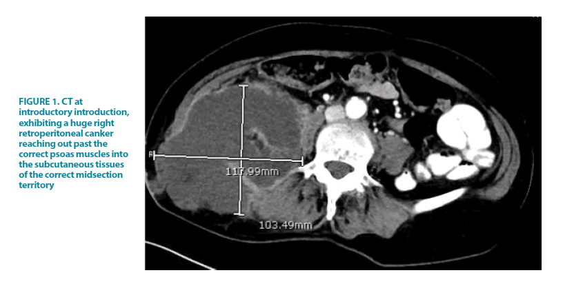
We report the unordinary instance of a 71-year-old female who had repetitive introductions more than three years with right midsection sore seeming to begin from a retroperitoneal assortment. She was at first determined to have a correct psoas canker of obscure etiology and was dealt with open careful waste of this assortment. Notwithstanding, further examinations after repetitive confirmations raised the doubt of an infected appendix as the hidden pathology. In spite of two unremarkable colonoscopies, she continued to go through analytic laparoscopy and appendectomy, with histopathology at last demonstrating interminable an infected appendix. She made an unremarkable recuperation.
Description
This portrays an uncommon sign of a typical pathology, with incessant an infected appendix introducing as a correct midsection ulcer with related cellulitis. An infected appendix, specifically retrocaecal an infected appendix, is an all-around perceived reason for psoas abscesses; anyway constant perceptible sore development of the midsection is uncommon and has not been portrayed in the writing. The analysis of a ruptured appendix ought to be considered in patients with no other away from of right flank boil or cellulitis related with a psoas ulcer.
There are not many cases in the writing of a ruptured appendix introducing as delicate tissue contaminations of the flank or stomach divider. Portrayed an instance of a retroperitoneal affixed boil introducing as right thigh cellulitis. For each situation, CT was basic in accomplishing the conclusion and right administration of the pathology. Be that as it may, this is the primary revealed instance of constant an infected appendix showing as a correct midsection ulcer with related cellulitis causing bleakness over numerous years [1].
References
Scali EP, Chandler TM, Heffernan EJ, et al. Primary retroperitoneal masses: What is the differential diagnosis? Abdom Imaging. 40, 1887-1903 (2015).
Lusuardi L, Kunit T, Janetschek G. Minimally Invasive Retroperitoneal Lymphadenectomy. J Endourol. 32, S97-S104 (2018).


