Case Report - Research on Chronic Diseases (2022) Volume 6, Issue 6
Neurofibromatosis emphysema and interstitial lung disease
Liu Yongjun*
Department of Radiology, Democritus University of Thrace, Greece
Department of Radiology, Democritus University of Thrace, Greece
E-mail: Yongjunlinedu123@gmail.com
Received: 01-Nov-2022, Manuscript No. OARCD-22-79064; Editor assigned: 03-Nov-2022, PreQC No. OARCD-22-79064 (PQ); Reviewed: 17-Nov-2022, QC No. OARCD-22-79064; Revised: 21-Nov-2022, Manuscript No. OARCD-22-79064 (R); Published: 30-Nov-2022, DOI: 10.37532/rcd.2022.6(6).123-125
Abstract
Lung cancer associated with neurofibromatosis type I is considered veritably rare, and only a many case reports have been described in the literature. There’s some substantiation that a inheritable relation between neurofibromatosis and carcinogenesis in the lung may live. We present a 42- time-old lady, continuance nonsmoker with a given history of neurofibromatosis type I, free of respiratory symptoms, which passed a low- cure HRCT of the lungs to probe any occult interstitial lung changes. A solitary ill- defined bump of ground- glass nebulosity was detected apropos in the middle lobe with no associated lymphadenopathy or metastatic complaint. Several thin- walled lung excrescencies were also seen in the lower lobes. Histological analysis of the bump after middle lobectomy revealed well- discerned adenocarcinoma. The case didn’t admit systemic chemotherapy or radiotherapy. She was free of complaint on 18- month follow up.
Introduction
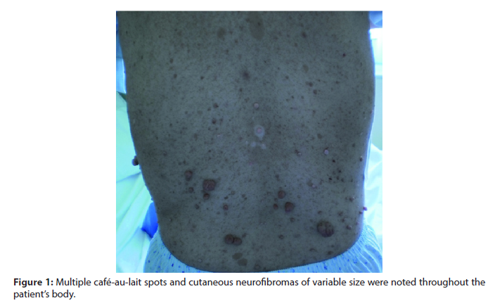
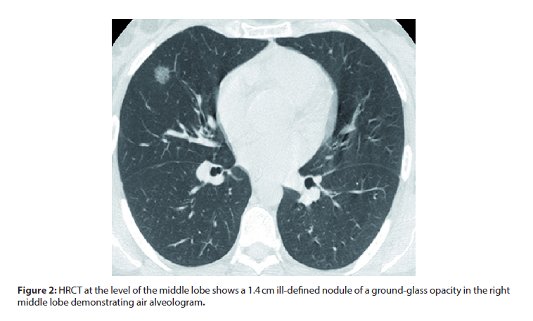
A 42- time-old lady, continuance nonsmoker with a given history of neurofibromatosis type I presented to the Outpatient Neurology Clinic for a regular follow- up. Physical examination revealed multiple cafe au- lait spots ´ and cutaneous neurofibromas of variable size throughout her body (Figure 1). She also suffered from vitiligo since the age of 20 times old. Her neurologic examination was normal, and she was free of respiratory symptoms. She reported no history of environmental or occupational exposure to other implicit carcinogens for lung cancer [1]. still, as part of her thorough follow- up, she passed a low- cure HRCT of the lungs to probe any occult interstitial lung abnormalities and presence of lung excrescencies that have been described in neurofibromatosis cases. HRCT of the lungs detected an incidental ill- defined solitary pulmonary bump1.4 cm in periphery, flaunting a ground- glass nebulosity and air alveologram (Figure 2). No other lung nodes or areas of connection were seen in any Case donation. A 42- time-old lady, continuance nonsmoker with a given history of neurofibromatosis presented to the Outpatient Neurology Clinic for a regular follow up [2]. Physical examination revealed multiple cafe au- lait spots ´ and cutaneous neurofibromas of variable size throughout her body (Figure 1). She also suffered from vitiligo since the age of 20 times old. Her neurologic examination was normal, and she was free of respiratory symptoms [3].
She reported no history of environmental or occupational exposure to other implicit carcinogens for lung cancer. still, as part of her thorough follow up, she passed a low- cure HRCT of the lungs to probe any occult interstitial lung abnormalities and presence of lung excrescencies that have been described in neurofibromatosis cases [4,5]. HRCT of the lungs detected an incidental ill- defined solitary pulmonary bump1.4 cm in periphery, flaunting a groundglass nebulosity and air alveologram (Figure 2) [6]. No other lung nodes or areas of connection were seen in any Discussion Neurofibromatosis type 1 or von Recklinghausen’s complaint is an autosomal dominant dysplasia of the ectoderm and mesoderm characterized substantially by the presence of neurofibromas, cafe- au- lait spots, and painted hamartomas ´ in the iris. The NF- 1 gene has been localized to chromosome 17q11 and functions as excrescence suppressor gene, and its separate gene product has been named as neurofibromin. It has been suspected that the mutation of excrescence suppressor NF- 1 gene increases the case’s threat for the development of colorful malice substantially deduced from the neural crest similar as nasty schwannoma, neurofibrosarcoma, intracranial glioma, and pheochromocytoma [7,8].
Still, the association of NF- 1 with lung cancer isn’t common. A review from Japan 20 times ago reported only 11 cases of NF- 1 with lung cancer, and we interestingly were suitable to find 7 further cases up to now- without including the presented case — also from the Japanese literature [9]. Adenocarcinoma was the most frequent histological opinion(72.9) in the first 11 cases reviewed, while the following 6 cases were diagnosed as no small cell cancer(3 cases), small cell cancer(1 case), inadequately discerned cancer (1 case), adenocarcinoma (1 case), and carcinosarcoma (1 case). Smoking habit wasn’t always mentioned in the reported cases in the literature, but there were some cases that were no way smokers as in our case [10,11]. In our case, histology revealed a welldiscerned adenocarcinoma, which is also the most common histological type of lung cancer in nonsmokers, who are by far more common in women. The case in the presented case was a continuance nonsmoker and so was her hubby. Since there’s no association with smoking in the presented case, a question raises whether there’s a inheritable relation between NF- 1 and the development of lung cancer [12]. NF- 1 gene has been counterplotted in the small part of chromosome 17q. p53 gene belongs to this area and has been intertwined in the development of colorful tumors including lung cancer. Also, in one study, it has been reported that the loss of heterozygosity in chromosome 17p — but not in 17q — was detected in a case with small cell melanoma with NF- 1 [13.14].
Conclusion
The authors hypothecated that the inactivation of excrescence suppressor gene on chromosome 17p — most likely p53 — might have been responsible for the development of small cell lung cancer in that case. Still, the association of lung cancer with NF- 1 might be coincidental. The bump in our case was of pure groundglass nebulosity which is known to have a high probability of malice compared to “solid nodes” and represents the “ Broncho alveolar ” element of lung adenocarcinoma indicating a better prognostic of lung adenocarcinoma. It remains to be answered whether the development of lung cancer in this youthful, no way - smoker, womanish case with NF- 1 simply followed the rules of the epidemiology of lung cancer in no way - smokers or there’s a inheritable link between NF- 1 and carcinogenesis in the lung. Large inheritable- grounded studies are demanded in order to probe these interesting findings. The evaluation of pulmonary involvement in NF- 1 cases with low- cure casket CT in long intervals could be proposed in order to descry early development of lung cancer or interstitial lung complaint.
Acknowledgement
None
Conflict of Interest
None
References
- Zamora AC, Collard HR, Wolters PJ et al. Neurofibromatosis-associated lung disease: a case series and literature review. Eur Clin Respir J. 29, 210-214 (2007).
- Muwakkit SA, Rodriguez Galindo C, Samra AI El et al. Primary malignant peripheral nerve sheath tumor of the lung in a young child without neurofibromatosis type 1. Pediatr Blood Cancer. 47, 636-638 (2006).
- Ginsberg RJ, Rubinstein LV. Randomized trial of lobectomy versus limited resection for T1 N0 non-small cell lung cancer: Lung Cancer Study Group. Ann Thorac Surg. 60, 615-623 (1995).
- Riccardi VM. Neurofibromatosis: past, present, and future. N Engl J Med. 324, 1283-1285 (1991).
- Ryu JH, Parambil JG, McGrann PS et al. Lack of evidence for an association between neurofibromatosis and pulmonary fibrosis. Chest. 128, 2381-2386 (2005).
- Itoi K, Yanagihara K, Okubo K et al. A case of lung cancer in a patient with von Recklinghausen’s disease. Nihon Geka Gakkai Zasshi. 30, 317-321(1992).
- Gupta KB, Kumar V, Tandon S et al. Primary carcinoma of the lung in von Recklinghausen neurofibromatosis. Lung India. 26, 130-132 (2009).
- Dikensoy O, Tunc ozg B. Primary adenocarcinoma of the lung in a case of von Recklinghausen neurofibromatosis. Turk J Med Sci. 32, 32-36 (2002).
- Fukuoka K, Katada H, Kohnoike Y et al. A case of von Recklinghausen’s disease associated with large cell carcinoma of the lung. Nihon Geka Gakkai Zasshi. 31, 88-93 (1993).
- Shimizu E, Shinohara T, Mori N et al. Loss of heterozygosity on chromosome arm 17p in small cell lung carcinomas, but not in neurofibromas, in a patient with von Recklinghausen neurofibromatosis. Cancer. 71, 725-728 (1993).
- Shimizu Y, Tsuchiya S, Watanabe S et al. Von Recklinghausen’s disease with lung cancer derived from the wall of emphysematous bullae. Intern Med. 33, 167-171 (1994).
- Satoh M, Wakabayashi O, Araya Y et al. Autopsy case of von Recklinghausen’s disease associated with lung cancer, gastrointestinal stromal tumor of the stomach, and duodenal carcinoid tumor. Nihon Kokyuki Gakkai Zasshi. 47, 798-804 (2009).
- Cıtıl R, Ciralik H, Karsligıl A et al. Carcinosarcoma of the lung associated with neurofibromatosis type 1: a case report. Turk Patoloji Derg. 28, 90-94 (2012).
- Foegle J, edelin GH, Lebitasy MP et al. Specific features of non-small cell lung cancer in women: a retrospective study of 1738 cases diagnosed in BasRhin between 1982 and 1997. J Thorac Oncol. 2, 466-474 (2007).
Indexed at, Crossref, Google Scholar
Indexed at, Crossref, Google Scholar
Indexed at, Crossref, Google Scholar
Indexed at, Crossref, Google Scholar
Indexed at, Crossref, Google Scholar
Indexed at, Crossref, Google Scholar
Indexed at, Crossref, Google Scholar
Indexed at, Crossref, Google Scholar
Indexed at, Crossref, Google Scholar


