Research Article - Clinical Investigation (2022) Volume 12, Issue 3
Application of simple imaging equipment based on smartphone camera function in diagnosis and treatment of eye diseases in Africa
- Corresponding Author:
- Ke Xiong
Department of Ophthalmology
Nanfang Hospital, Southern Medical University, Guangzhou, China
E-mail: shinco1223@163.com
Received: 11-March-2022, Manuscript No. fmci-22-56863; Editor assigned: 13-March-2022, PreQC No. fmci-22-56863 (PQ); Reviewed: 22-March-2022, QC No. fmci-22-56863 (Q); Revised: 23-March-2022, Manuscript No. fmci-22-56863 (R); Published: 31-March-2022; Doi: 10.37532/2041-6792.2022.12(3).69-74
Abstract
The eye image data is very important in the recording of ophthalmic diseases. Various imaging equipments, such as anterior segment camera, fundus-imaging machines, optical coherence tomography scanner, are very expensive and cumbersome. Only a few eye clinics in most African countries have these equipments, the most eye clinics are only equipped with basic ophthalmic examination equipment such as slit-lamp microscope and ophthalmoscope. Now, the miniature camera of smartphones has the ability to take high-resolution photographic images. Combining the smartphone's camera functions and auxiliary simple devices connected to mobile phones can collect eye imagings for clinical applications. The author introduced a simple and cheap device for eye imaging. The method of combining the camera function of a smartphone with a simple imaging device to collect images of the anterior segment and the fundus. It’s simple, easy-to-use and low-cost characteristics can fill this shortcoming in Africa. It can be more familiar and mastered by African ophthalmologists.
Keywords
smartphone • Anterior segment photography • Fundus photography • Eye clinic • Africa
Introduction
The collection of ophthalmological image data is very important in the diagnosis and recording of ophthalmic diseases [1-2]. At present, the eye clinics in developed countries are equipped with various imaging equipment such as anterior segment camera, fundus-imaging machines, optical coherence tomography scanner and so on. These instruments are very expensive, unaffordable, and require specialized personnel. Due to economic backwardness and limited funds, the most eye clinics in African countries are only equipped with basic ophthalmic examination equipment such as slit-lamp microscope and ophthalmoscope. With the development of technology and the popularization of mobile phones, the miniature camera of smartphones has the ability to take high-resolution photographic images. Combining the smartphone's camera functions and auxiliary simple devices connected to mobile phones can collect eye imagings for clinical applications [3-4]. The author as ophthalmologist who is dispatched by Chinese government and worked a suburban hospital in Ghana, which is a low-income country in West Africa. He introduced a simple and cheap device for eye imaging that applied the smartphone's camera function with simple imaging equipment to collect anterior segment and fundus image.
Methods and Materials
Selection of phone
This study used iPhone7, which uses the Apple A10 Fusion quad-core processor. The rear single camera has a 12-megapixel image sensor, automatic image stabilization and image stabilization, f/1.8 aperture, Focus Pixels autofocus and tap-to-focus, continuous shooting and noise reduction, and automatic HDR photos. iPhone7 also supports slow motion and 4K video recording. The iPhone7 model is thin and light, and the rear single camera is easier to match with the connected device [5].
Selection of device
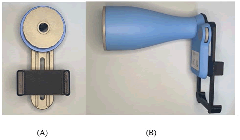
The slit lamp microscope without camera functions can be used. Slit lamp mobile phone holder (Figure 1A) is bought from China, that consists of two parts: mobile phone fixed slot and slit lamp eyepiece slot. Suitable for all kinds of smartphones with camera function, the price is only about $100. Indirect ophthalmoscope (Figure 1B) is hand-held and portable, using the working principle of indirect ophthalmoscope, after the examinee dilated, the examiner uses a smartphone to record the fundus of the patient's eye through the ophthalmoscope and observe fundus video and HD images are saved. It is suitable for all kinds of smartphones with camera functions,but the phone slot needs to be customized. It is also bought from China, and the price is just about $400.
Anterior segment photography
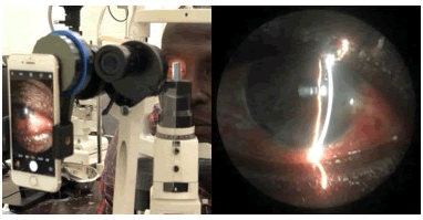
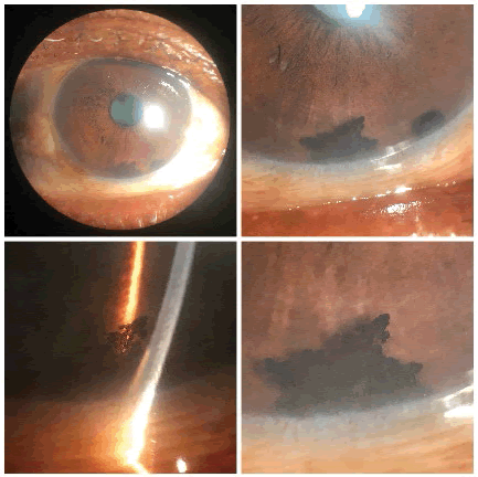
First fix the mobile phone to the mobile phone slot of the holder, rotate the mobile phone to align the camera on the back of the mobile phone with the camera hole, and connect the holder to the left eyepiece of the slit lamp, which is easy to operate. Rotating the bracket button fixes the slit lamp eyepiece slot, and the mobile phone screen is kept vertical, ensuring that the two optical systems of the slit lamp microscope and camera can fully match each other. During the video recording process, adjust the seat and the slit lamp to be in a comfortable position. Operation in a relatively dark room can eliminate clutter reflections that interfere with the light source. The light source selects the standard viewing illumination on a slit lamp without the need for a mobile phone's flash. The light intensity and projection angle of the slit lamp can be adjusted as needed, and the slit length is appropriate to reduce redundant reflections (Figure 2). Reasonable selection of diffuse light source and slit light band photography, the former can show the general profile of the anterior segment of the eye, and the latter can highlight the structural levels and details of the anterior segment of the eye. You can use an additional flashlight to make auxiliary background lighting, which can illuminate the background tissue to make the image more saturated and delicate, and better reflect the close anatomy of the narrow slit optical section. The optical zoom of the slit lamp and the digital zoom of the mobile phone can be adjusted to adjust the magnification, but a high digital zoom will reduce the image resolution, increase the graininess, and the optical zoom affects the depth of field. When it is really necessary to zoom in for further observation of local tissue, optical zoom is still preferred. The image can also be cropped later to highlight the target (Figure 3). Similarly, a slit lamp can be used in combination with gonioscope and Hruby preset lens to take pictures of the corner and fundus. After a period of practice of techniques, the sharpness, chroma, and contrast of the collected photos are satisfactory, and the tissue structure and lesions can be clearly displayed [6].
Fundus photography
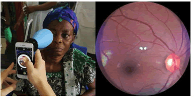
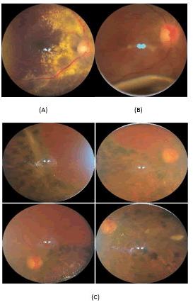
Although the slit lamp combined with Hruby preset lens can be used for fundus photography, the scope of the fundus is small, and the operation of preset lens inspection and mobile phone photography is difficult. We choose the indirect ophthalmoscope. After the examinee is dilated, fix the mobile phone in the slot of the instrument, turn on the light source of the device, and move the ophthalmoscope lens to the examinee, about 5 cm away from the cornea. Although the mobile phone has optical image stabilization, it is selected in the video capture mode of the mobile phone camera. When shooting, you need to stabilize the device with both hands and a moderate shift to obtain a comprehensive fundus photo. It takes several attempts to grasp Best focus and image quality, and movement is in the opposite direction. Through the movement of the device and the autofocus function of the mobile phone, a comprehensive dynamic video recording of the fundus can be performed, and the fundus range can be seen more than 50°. After capturing the continuous illumination video through the light source that comes with the device, select a specific frame as the image representing the fundus (Figure 4). Compared with the fundus photograph under the preset lens, the field of view is large, the resolution is well, and the image is sufficient for document recording [7].
Results
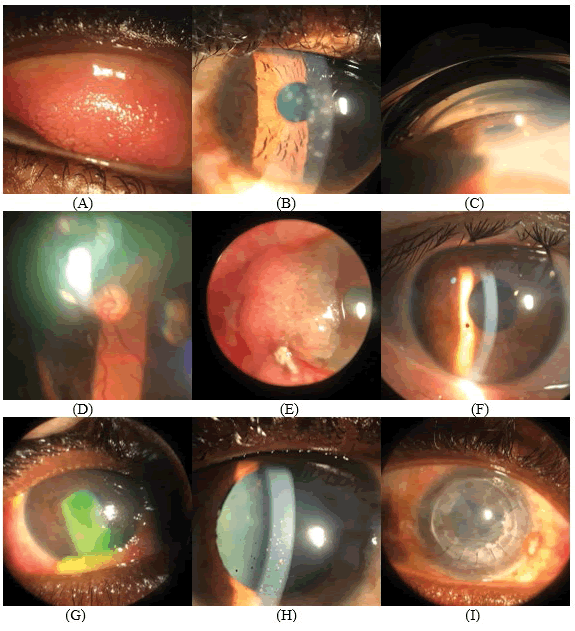
A large number of anterior segment and fundus photos provided by the simple imaging device. The sharpness and color of these collected photos can reflect the lesions of the eye (Figure 5 and Figure 6).
The photos taken were taken by the author. The phone has excellent imaging capabilities and shows excellent features. Although the quality of imaging provided by mobile phones is lower than that of professional cameras and equipments, it is sufficient for recording and diagnosis. Photos are made through the simple imaging devices such as the slit lamp mobile phone holder and indirect ophthalmoscope, which makes the mobile phone camera very easy to perform. After repeated attempts over time and time, you can master this method. Closely cooperate with the proficiency of the operation to obtain higher quality anterior segment and fundus photos. The collected images can be edited, Organized and archived through the photo editing function of the mobile phone, which can record the changes of the patient's condition. These images greatly facilitate the research and learning of diseases; at the same time, they can be sent to patients through social APP such as WhatsApp, WeChat, and also facilitate communication with local patients; remote consultation for difficult cases can improve the accuracy of disease diagnosis; Communication between peers through academic platforms is conducive to joint learning and improvement.
Discussion
High-quality ophthalmic imaging data have very important clinical value in the diagnosis and recording of ophthalmic diseases [1]. Owing to the economic backwardness and lack of funds in Africa, such as Ghana, it is not possible to purchase expensive eye imaging equipment such as fundus camera and anterior segment camera all eye clinics. Most eye clinics are generally equipped only with slit lamp microscope and direct fundus cope [8]. With the advancement of technology, mobile phones have become an indispensable, and high-resolution digital high-definition camera has become the default function of smartphones, making mobile phones a powerful tool for ophthalmic imaging. The method of combining the camera function of a smartphone with a simple imaging device to collect images of the anterior segment and the fundus. It’s simple, easy-to-use and low-cost characteristics make up for the shortcomings of obtaining image data and can fill this shortcoming in the vast region of Africa [9].
Slit lamp mobile phone holder is suitable for all kinds of smartphones with camera function, and all kinds of slit lamp microscope models can be used. Indirect ophthalmoscope uses the working principle of indirect ophthalmoscope, which requires dilated pupil inspection. It is suitable for all types of smartphones with camera functions, but the phone slot needs to be customized. Both devices are simple, portable, and inexpensive, making them suitable for use in Africa where advanced and expensive imaging technology and equipment cannot be afforded [10,11]. At present, the smartphone is very powerful and has the function of comprehensive information processing. Its convenience and operability can be widely used in clinical work. Smartphones provide high-quality anterior segment and fundus photos, and the quality of the captured images can better meet the clinical needs. Smartphones also allow third-party applications to customize the user experience, such as vision inspection, color blindness inspection, and so on, which can comprehensively inspect and evaluate visual function and eye conditions, and it also obtain relevant medical information. [1,5]
Connect your smartphone with a simple hardware device without the need for a third-party application. Clinicians can master this technique quickly and repeatedly after repeated exercises, and the quality of the captured images can better meet the clinical needs. An easy-to-connect device is used to take pictures of the anterior segment of the eye and the fundus, and obtain images for intuitive communication, making it easier for patients to understand the condition. With the images, it is possible to explain the problem to patients and perform timely treatment. For the images of difficult cases, telemedicine consultations in advanced hospitals can be performed to improve the accuracy of disease diagnosis; it is also convenient for academic exchanges and learning among peers [2].
Conclusion
The low-cost anterior segment and fundus inspection imaging technology of the mobile phone camera function completely eliminate the hassle of purchasing large eye camera equipment. The quality of the captured images can better meet the clinical needs, and it can also be accessed through the mobile phone network function for data transmission, archiving and communication. This method can be used as a routine inspection and recording method, can better improve the objective data of cases, can be popularized in Africa and can be more familiar and mastered by African ophthalmologists.
Conflict of interest
The authors have no relevant financial or non-financial interests to disclose.
Reference
- Li HK. Telemedicine and ophthalmology. Surv Ophthalmol. 44:61-72 (1999).
Google Scholar Crossref - Houts PS, Doak CC, Doak LG, et al. The role of pictures in improving health communication: A review of research on attention, comprehension, recall, and adherence. Patient Educ Couns. 61:173-190 (2006).
Google Scholar Crossref - Lord RK, Shah VA, San Filippo AN, et al. Novel uses of smartphones in ophthalmology. Ophthalmology. 117:1274-1274.e3 (2010).
Google Scholar Crossref - Kim DY, Delori F, Mukai S. Smartphone photography safety. Ophthalmology. 119:2200-2201 (2012).
Google Scholar Crossref - Jalil M, Ferenczy SR, Shields CL. iPhone 4s and iPhone 5s Imaging of the Eye. Ocul Oncol Pathol. 3:49-55 (2017).
Google Scholar Crossref - Hester C. Slit-lamp photography with a smartphone. Adv Ocular Care. 2012:62-63 (2012).
Google Scholar Crossref - Bastawrous A. Smartphone fundoscopy. Ophthalmology. 119:432-433 (2012).
Google Scholar Crossref - Idriss BR, Tran TM, Atwine D, et al. Smartphone-based ophthalmic imaging compared with spectral-domain optical coherence tomography assessment of vertical cup-to-disc ratio among adults in Southwestern Uganda. J Glaucoma. 30:e90-e98 (2021).
Google Scholar Crossref - Mohammadpour M, Heidari Z, Mirghorbani M, et al. Smartphones, tele-ophthalmology, and VISION 2020. Int J Ophthalmol. 10:1909-1918 (2017).
Google Scholar Crossref - Diethei D, Schöning J. Using smartphones to take eye images for disease diagnosis in developing countries. In Proceedings of the Second African Conference for Human Computer Interaction: Thriving Communities. 2018:1-3 (2018).
Google Scholar Crossref - Wintergerst MW, Jansen LG, Holz FG, et al. Smartphone-based fundus imaging-Where are we now? Asia-Pacific J Ophthalmol. 9:308-314 (2020).
Google Scholar Crossref
