Case Report - Clinical Practice (2017) Volume 14, Issue 1
A longitudinal study of angular artery island flap, used for reconstruction of facial defects
- Corresponding Author:
- Arghya Bera
RGKAR Medical College Kolkata
West Bengal India
E-mail: drarghyabera7@gmail.com
Abstract
Considering cosmetic and functional outcome, reconstruction of moderate to large mid & upper facial soft-tissue defects due to trauma, neoplasm, or infection remains a challenge. We used either ipsilateral or contralateral angular artery island flap in patients with full-thickness soft-tissue defects in those areas. We present our experience in 30 patients (17 females & 13 males) with mean age of 65 , with complex soft-tissue defects in mid & upper face reconstructed with angular artery island flaps. Defect sizes changed from 1 × 2 cm to3.5 × 5 cm. Flap size varied from a length of 2.2 to 6 cm average (average 4 cm) and width of 2.7 to 6.5 cm (average 5 cm). All donor sites were closed primarily. Twenty seven flaps (90%) healed without any necrosis and completely survived. Ipsilateral or contralateral angular artery island flap is a very convenient, safe and reliable flap for reconstruction of moderate to large mid and upper facial defects. Good aesthetic outcome for variety of facial defects could be obtained with this flap. Donor site morbidity also less.
Keywords
mid and upper facial defect, angular artery island flap, ipsilateral or contralateral, local flap, donor area
Introduction
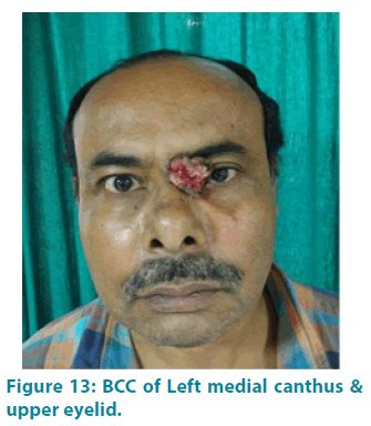
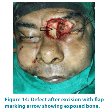
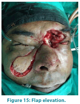
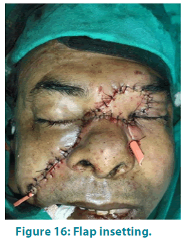
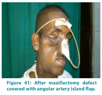
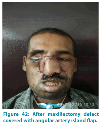
Considering cosmetic and functional outcome, reconstruction of moderate to large mid & upper facial soft-tissue defects due to trauma, neoplasm, or infection remains a challenge. We used either ipsilateral or contralateral angular artery island flap in patients with full-thickness soft-tissue defects in those areas.
We present our experience in 30 patients (17 females & 13 males) with mean age of 65 , with complex soft-tissue defects in mid & upper face reconstructed with angular artery island flaps. Defect sizes changed from 1 × 2 cm to3.5 × 5 cm.
Flap size varied from a length of 2.2 to 6 cm average (average 4 cm) and width of 2.7 to 6.5 cm (average 5 cm). All donor sites were closed primarily. Twenty seven flaps (90%) healed without any necrosis and completely survived.
Ipsilateral or contralateral angular artery island flap is a very convenient, safe and reliable flap for reconstruction of moderate to large mid and upper facial defects. Good aesthetic outcome for variety of facial defects could be obtained with this flap. Donor site morbidity also less.
| Age range (years) | Number |
|---|---|
| 31-40 | 1 |
| 41-50 | 2 |
| 51-60 | 6 |
| 61-70 | 9 |
| 71-80 | 9 |
| >80 | 3 |
Table 1: Age distribution.
| Sex | Number |
|---|---|
| Male | 13 |
| Female | 17 |
Table 2: Sex distribution
| Diabetes | Number |
|---|---|
| Yes | 9 |
| No | 21 |
Table 3: Diabetes.
| Lesion | Number |
|---|---|
| BCC | 18 |
| SCC | 12 |
Table 4: Nature of lesion.
| Site | Number |
|---|---|
| Paranasal | 1 |
| Infra orbital | 6 |
| Medial canthus | 8 |
| Malar region | 6 |
| Nasal dorsum | 4 |
| Nasal tip | 2 |
| Glabella | 1 |
| Nasal ala | 2 |
Table 5: Site of lesions.
| Length (cm) | Breadth (cm) | ||
|---|---|---|---|
| Minimum | Maximum | Minimum | Maximum |
| 1 | 4.5 | 1.5 | 5 |
Table 6: Size of lesions.
| Length (cm) | Breadth (cm) | ||
|---|---|---|---|
| Minimum | Maximum | Minimum | Maximum |
| 2 | 5.5 | 2 | 6 |
Table 7: Size of defects.
| Length (cm) | Breadth (cm) | ||
|---|---|---|---|
| Minimum | Maximum | Minimum | Maximum |
| 2.2 | 6 | 2.7 | 6.5 |
Table 8: Size of flaps.
| Maximum (mins) | Minimum (mins) | Mean |
|---|---|---|
| 120 | 70 | 89.17 |
Table 9: Operative time-time required to create defect, raise flap & inset of flap.
| Maximum (days) | Minimum (days) | Mean |
|---|---|---|
| 27 | 6 | 12.50 |
Table 10: Post-operative hospital stay.
| Complication | Number |
|---|---|
| No complication | 23 |
| Partial necrosis | 3 |
| Bulky | 2 |
| Haematoma | 1 |
| Ectropion | 1 |
Table 11: Complication.
| Smoking | Complication | No complication | p value |
|---|---|---|---|
| Yes | 3 | 8 | 0.698 |
| No | 4 | 15 |
Table 12: Relation between flap survival and smoking.
| Diabetes | Complication | No complication | p value |
|---|---|---|---|
| Yes | 3 | 6 | .397 |
| No | 4 | 17 |
Table 13: Relation between flap survival and Diabetes.
| Null Hypothesis | Test | Sig. | Decision |
|---|---|---|---|
| The distribution of Flap_L is the same across categories of Complication_New. |
Independent-Samples Mann-Whitney U Test |
0.0541 | Retain the null hypothesis. |
| The distribution of Flap_B is the same across categories of Complication_New. |
Independent-Samples Mann-Whitney U Test |
0.0271 | Retain the null hypothesis. |
| Asymptotic significances are displayed. The significance level is 0.05. 1Exact significance is displayed for this test. |
|||
Table 14: Relation between flap dimension and complication
| Best outcome | Worst outcome | Mean outcome |
|---|---|---|
| 6 | 14 | 8.6 |
Table 15: Facial aesthetics.
