Commentary - Clinical Investigation (2022) Volume 12, Issue 8
Reverse Engineering Techniques on Musculoskeletal System
- Corresponding Author:
- Shenxing Du
Department of Rehabilitation, Dongyang People’s Hospital, Dongyang City, Zhejiang Province, China
E-mail: dsx0221@gmail.com
Received: 21-July-2022, Manuscript No. fmci-22-69892; Editor assigned: 23-July-2022, PreQC No. fmci-22-69892 (PQ); Reviewed: 03-August-2022, QC No. fmci-22-69892 (Q); Revised: 05-August-2022, Manuscript No. fmci-22-69892 (R); Published: 15-August-2022, DOI: 10.37532/2041-6792.2022.12(8).146-149
Abstract
Commentary
Some symptoms have nothing to do with injuries, including pain, let alone those situations where there is no injury. It was very occasional for us to get in touch with Reverse Engineering Techniques (RETs) while we were evaluating and treating some very special cases which had no abnormal signs and few improvements based on routine treatments. Most RETs are difficult to accurately determine three-Dimensional (3D) bone motion in vivo, especially the subtle differences of the intraarticular relationship, due to the unreliability or misery to directly attach skin or bone-mounted markers [1]. Additionally, the bone shape which is the chief culprit of abnormal mobility varies in each body, although humans have “same” structures.
3D to 2D Image Registration Technique (3D-2D IRT) can help us attain easy access to the in-vivo measurement of the joint dynamic motions using their own bones while avoiding using available cadaveric experimental data and analyze the intraarticular motions [2]. I am obsessed with this technique from the first contact, not only because it helps us evaluate subtle activities of each joint, but because it allows us to intuitively understand the laws of intraarticular motion, thus it becomes an indispensable part of my clinical teaching programs. Furthermore, it can guide treatments, such as the directions and degrees of dynamic fixation via rigid tape [2,3]. By means of collaborating with orthopedists, they can also apply this technique for surgical evaluation and patient education.
However, according to our practice, 3D-2D IRT has two main drawbacks.
Firstly, it just involves the 3D dynamic processing and position quantitation. So, we must use math models to quantitate the process of dynamic motion of each bone. Continuous Transformation Matrix (CTM) and Dual Quaternion (DQ) were in sight [4]. We applied the CTM only in our previous article due to the intuitive figures and norms. All the Bone Coordinate System (BCS) data are relative to the World Coordinate System (WCS), so we recommend transforming the data to set a relative stable bone’s BCS as a new WCS for comparison. The DQ describes the rotation and translation between CSs. In terms of dynamic motion quantitation, it would be better than the CTM. But it is not intuitive enough for beginners or easy for those who are not good at advanced mathematics.
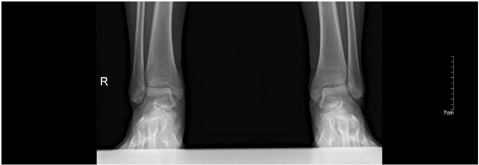
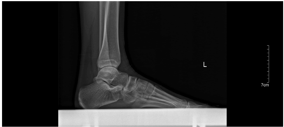
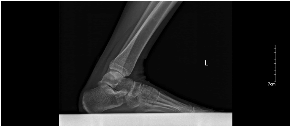
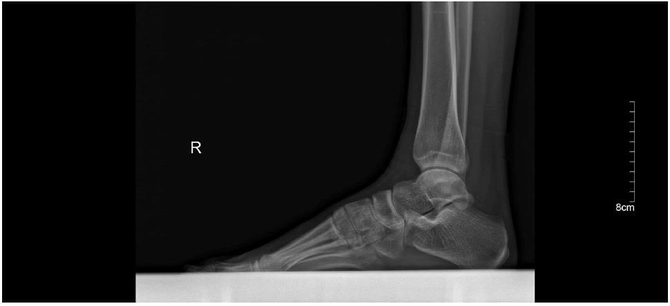
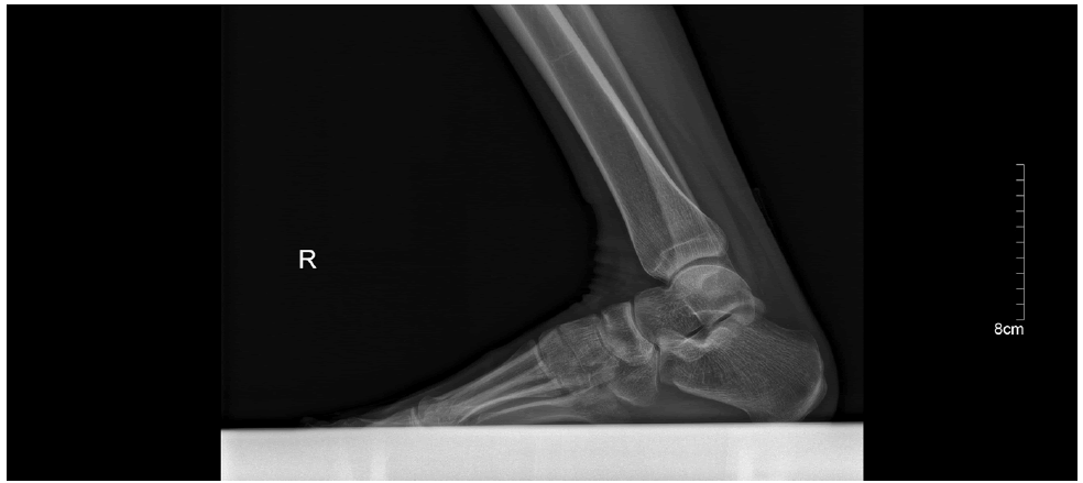
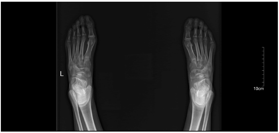
Secondly, the processing is cumbersome. Even a skilled practitioner would take at least three days to complete all analyses of a patient. So, we simplified the processing via dimension reduction. We designed a new X-ray photography. Against ankle, it includes 6 under weight-bearing (Figure 1-6) and 1 under non-weight-bearing (Figure 7). It is easy for us to unscramble most subtle activities based on this kind of X-ray photography. Therefore, in clinical practice we apply 3D-2D IRT only on the high rate of variation of bone shape due to computed tomography or musculoskeletal problems for which the cause cannot be identified. Furthermore, 3D2D IRT should combine math models for scientific research.
In summary, RETs can be a good way for complicated non-injured musculoskeletal issues, especially for subtle activities. Besides, RETs should be modified based on clinical and research requirements.
This manuscript is a short commentary of the article “Subtle Activities of Specific Plain Subtalar Joint May Account for Non-injured Ankle Pain: A Case Report” [2].
References
- Dennis DA, Mahfouz MR, Komistek RD, et al. In vivo determination of normal and anterior cruciate ligament- deficient knee kinematics. J Biomech. 38(2):241-253 (2005).
- Du S, Wei L. Subtle Activities of Specific Plain Subtalar Joint May Account for Non-injured Ankle Pain: A Case Report. Int J Biomed Eng Clin Sci. 7(4):91-95 (2021).
- Du S, Wei L, He B. Dynamic fixation using rigid tape in rehabilitation after surgery of terrible triad injury of the elbow: A randomized trial. J Back Musculoskelet Rehabil. 34(6):957-964 (2021).
- Wu YX, Hu XP, Hu DW, et al. Strapdown inertial navigation system algorithms based on dual quaternions. IEEE Trans Aerosp Electron Syst. 41(1):110-132 (2005).

