Clinical images - Imaging in Medicine (2017) Volume 9, Issue 5
Large defect on lung scintigraphy mimicking pulmonary embolism
Aparna Komarraju, Tracy L Yarbrough, Jorge Brito, Twyla Bartel*Global Advanced Imaging, PLLC Little Rock, AR, USA
- Corresponding Author:
- Twyla Bartel
Global Advanced Imaging
PLLC Little Rock, AR, USA
E-mail: twylabb@hotmail.com
Abstract
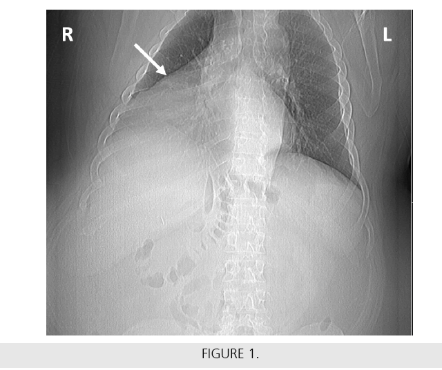
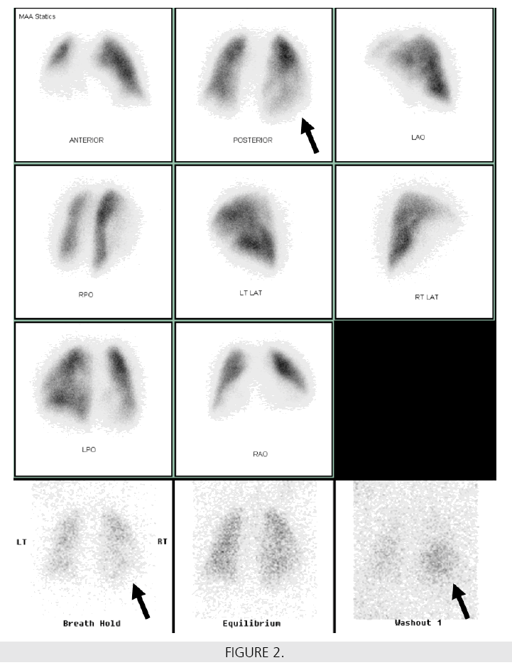
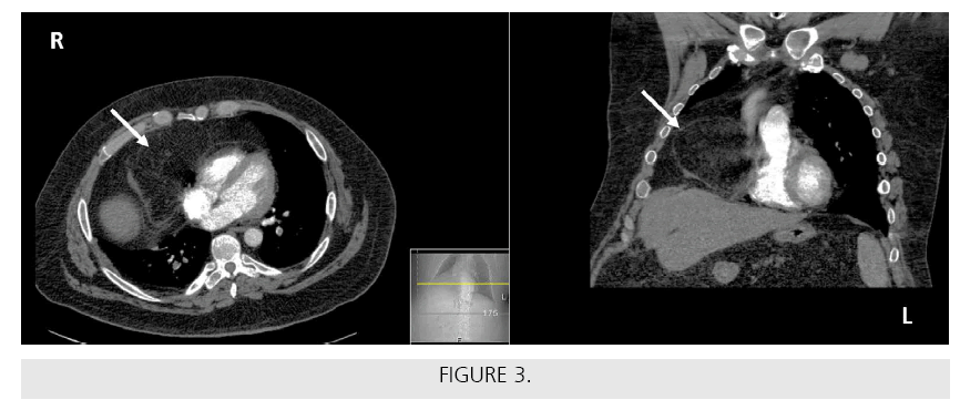
A middle-aged patient with acute shortness of breath had negative cardiac/GI workups. Chest x-ray showed mild atelectasis and a large opacification silhouetting the right heart border/ hemidiaphragm (FIGURE 1). A subsequent lung (VQ) scan (99mTc-MAA perfusion-top 3 rows; 133Xe ventilation-last row) showed a large right lower lung triple match and was interpreted as intermediate probability for pulmonary embolism (PE) (FIGURE 2). Also note the radiopharmaceutical retention in the area of concern in the right lower lung on the ventilation washout image shown (last row, last image to the right). Symptoms continued despite anticoagulation. A contrast-enhanced chest CT was then performed showing a large Morgagni hernia in the right lung (FIGURE 3).
This was, in fact, the cause of the patient's shortness of breath rather than PE. A Morgagni hernia is congenital, is typically located in the anterior chest, and is more common on the right side (as seen on these images). A patient with Morgagni hernia may not become symptomatic until adulthood. The retention of the xenon radiopharmaceutical was likely due to longterm obstruction to this region from the large hernia with trapping of 133Xe. An alternate proposal for 133Xe retention in the area of this hernia on the ventilation views was due to the fat content of the herniated omental fat as it is known that 133Xe tends to concentrate more in fat (i.e., well known to accumulate in a fatty liver). This is the first to our knowledge where a large Morgagi hernia caused the appearance of a triple match on VQ scintigraphy. If a SPECT/ CT camera had been available, it would have been very useful to simply further acquire SPECT/CT images immediately to determine the cause of the unusual image pattern in the right lower lobe while the patient was still in the Nuclear Medicine department.


